Download
1 / 17
170 likes | 472 Views
Chapter 8b. Neurons: Cellular and Network Properties. Axon of presynaptic neuron. Mitochondrion. Axon terminal. Postsynaptic neuron. Synaptic vesicles. Synaptic cleft. Neurotransmitter. Receptors. Postsynaptic membrane. Cell-to-Cell: A Chemical Synapse.

E N D
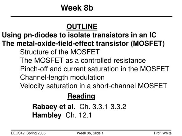
Chapter 8b Neurons: Cellular and Network Properties
Axon ofpresynapticneuron Mitochondrion Axon terminal Postsynaptic neuron Synapticvesicles Synapticcleft Neurotransmitter Receptors Postsynapticmembrane Cell-to-Cell: A Chemical Synapse • Chemical synapses use neurotransmitters; electrical synapses pass electrical signals. Figure 8-20
Cell-to-Cell: Events at the Synapse and Exocytosis 1 An action potential depolarizes the axon terminal. 2 Axon terminal The depolarization opens voltage-gated Ca2+ channels and Ca2+ enters the cell. Synaptic vesicle Neurotransmitter molecules 3 Calcium entry triggers exocytosis of synaptic vesicle contents. Action potential 4 Neurotransmitter diffuses across the synaptic cleft and binds with receptors on the postsynaptic cell. 1 3 Ca2+ 5 Neurotransmitter binding initiates a response in the postsynaptic cell. Synaptic cleft Docking protein 2 4 Receptor Voltage-gated Ca2+ channel Postsynaptic cell Cell response 5 Figure 8-21
Cell-to-Cell: Neurocrines • Seven classes by structure • Acetylcholine • Amines • Amino acids • Purines • Gases • Peptides • Lipids
Cell-to-Cell: Synthesis and Recycling of Acetylcholine at a Synapse Myasthenia gravis Mitochondrion Axon terminal CoA Acetyl CoA Acetylcholine Enzyme 1 1 Acetylcholine (ACh) is made from choline and acetyl CoA. Synaptic vesicle 2 In the synaptic cleft ACh is rapidly broken down by the enzyme acetylcholinesterase. 3 Choline Cholinergic receptor 2 3 Choline is transported back into the axon terminal and is used to make more ACh. Postsynaptic cell Acetate Acetylcholinesterase (AChE) Figure 8-22
Amines • Derived from single amino acid • Tyrosine • Dopamine • Norepinephrine is secreted by noradrenergic neurons • Epinephrine • Others • Serotonin is made from tryptophan • Histamine is made from histadine
Amino Acids • Glutamate: Excitatory CNS • Aspartate: Excitatory brain • GABA: Inhibitory brain • Glycine • Inhibitory spinal cord • May also be excitatory
Other Neurotransmitters • Purines • AMP and ATP • Gases • NO and CO • Peptides • Substance P and opioid peptides • Lipids • Eicosanoids
Receptors • Cholinergic receptors • Nicotinic on skeletal muscle, in PNS and CNS • Monovalentcation channels Na+ and K+ • Muscarinicin CNS and Parsympathetic NS • Linked to G proteins to 2nd messengers • Adrenergic Receptors • and • Linked to G proteins and 2nd messengers • Glutaminergic • Excitatory in CNS • Metabotropic and Ionotropic
Presynaptic axon terminal Slow synaptic potentials and long-term effects Rapid, short-acting fast synaptic potential Neurocrine G protein–coupled receptor Chemically gated ion channel Inactive pathway Postsynaptic cell Alters open state of ion channels Activated second messenger pathway Modifies existing proteins or regulates synthesis of new proteins Ion channels open Ion channels close More K+ out or Cl– in Less K+ out More Na+ in Less Na+ in IPSP = inhibitory hyperpolarization EPSP = excitatory depolarization EPSP = excitatory depolarization Coordinated intracellular response Cell-to-Cell: Postsynaptic Response • Fast and slow responses in postsynaptic cells Figure 8-23
Cell-to-Cell: Postsynaptic Response Presynaptic axon terminal Rapid, short-acting fast synaptic potential Neurocrine G protein–coupled receptor Chemically gated ion channel Postsynaptic cell Ion channels open More K+ out or Cl– in More Na+ in IPSP = inhibitory hyperpolarization EPSP = excitatory depolarization Figure 8-23, step 1
Cell-to-Cell: Postsynaptic Response Presynaptic axon terminal Slow synaptic potentials and long-term effects Rapid, short-acting fast synaptic potential Neurocrine G protein–coupled receptor Chemically gated ion channel Postsynaptic cell Ion channels open More K+ out or Cl– in More Na+ in IPSP = inhibitory hyperpolarization EPSP = excitatory depolarization Figure 8-23, steps 1–2
Cell-to-Cell: Postsynaptic Response Presynaptic axon terminal Slow synaptic potentials and long-term effects Rapid, short-acting fast synaptic potential Neurocrine G protein–coupled receptor Chemically gated ion channel Inactive pathway Postsynaptic cell Alters open state of ion channels Activated second messenger pathway Ion channels open More K+ out or Cl– in More Na+ in IPSP = inhibitory hyperpolarization EPSP = excitatory depolarization Figure 8-23, steps 1–3
Cell-to-Cell: Postsynaptic Response Presynaptic axon terminal Slow synaptic potentials and long-term effects Rapid, short-acting fast synaptic potential Neurocrine G protein–coupled receptor Chemically gated ion channel Inactive pathway Postsynaptic cell Alters open state of ion channels Activated second messenger pathway Ion channels open Ion channels close More K+ out or Cl– in Less K+ out More Na+ in Less Na+ in IPSP = inhibitory hyperpolarization EPSP = excitatory depolarization Figure 8-23, steps 1–4
Cell-to-Cell: Postsynaptic Response Presynaptic axon terminal Slow synaptic potentials and long-term effects Rapid, short-acting fast synaptic potential Neurocrine G protein–coupled receptor Chemically gated ion channel Inactive pathway Postsynaptic cell Alters open state of ion channels Activated second messenger pathway Ion channels open Ion channels close More K+ out or Cl– in Less K+ out More Na+ in Less Na+ in IPSP = inhibitory hyperpolarization EPSP = excitatory depolarization EPSP = excitatory depolarization Figure 8-23, steps 1–5
Cell-to-Cell: Postsynaptic Response Presynaptic axon terminal Slow synaptic potentials and long-term effects Rapid, short-acting fast synaptic potential Neurocrine G protein–coupled receptor Chemically gated ion channel Inactive pathway Postsynaptic cell Alters open state of ion channels Activated second messenger pathway Modifies existing proteins or regulates synthesis of new proteins Ion channels open Ion channels close More K+ out or Cl– in Less K+ out More Na+ in Less Na+ in IPSP = inhibitory hyperpolarization EPSP = excitatory depolarization EPSP = excitatory depolarization Coordinated intracellular response Figure 8-23, steps 1–6
Cell-to-Cell: Inactivation of Neurotransmitters 1 Neurotransmitters can be returned to axon terminals for reuse or transported into glial cells. Rapid termination of NTs 2 Enzymes inactivate neurotransmitters. Blood vessel 3 Neurotransmitters can diffuse out of the synaptic cleft. Axon terminal of presynaptic cell Synaptic vesicle 3 Glial cell 1 Enzyme 2 Postsynaptic cell Figure 8-24