Download
1 / 41
410 likes | 431 Views
Digestive System. FUNCTIONS. Ingestion Secretion (release of water, acid, buffers & enzymes into the GI tract) Mixing & propulsion Digestion 1. Chemical 2. Mechanical Absorption from the GI tract into the blood & lymph. Functions continued. Defecation. OVERVEIW. GI Tract

E N D
FUNCTIONS • Ingestion • Secretion (release of water, acid, buffers & enzymes into the GI tract) • Mixing & propulsion • Digestion 1. Chemical 2. Mechanical • Absorption from the GI tract into the blood & lymph
Functions continued • Defecation
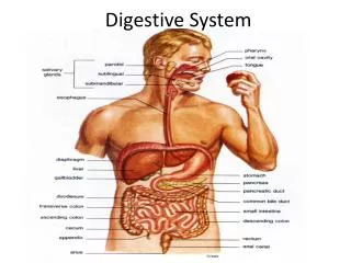
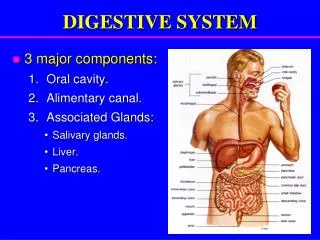
OVERVEIW • GI Tract 1. Mouth 2. Pharynx 3. Esophagus 4. Stomach 5. Large & small intestine • Accessory digestive organs • Teeth • Tongue
ACCESSORY DIGESTIVE ORGANS 3. Salivary glands 4. Liver 5. Gallbladder 6. Pancreas
LAYERS OF THE GI TRACT • Mucosa • 1. Mucus membrane that lines the lumen of the GI tract • 2. Mouth, pharynx & esophagus contains nonkeritanized stratified squamous epithelium • 3. Simple columnar lines stomach and intestine • 4. contains exocrine cells that secrete mucus • 5. Lamina propria • A, Aerolar connective Tissue • B. Contains lymphatic & blood vessels for nutrient absorption
Layers of the GI Tract Continued • C. Contains mucosa associated lymphoid tissue (MALT) that houses cells that protect against disease 6. Musculairs mucosae • A. Thin layer of smooth muscle • B. Causes folds that increase surface area • Submucosa • 1. Consists of areolar tissue • 2. Contains submuscosal plexus • A. Regulates movement of muscosa & vasoconstriction of blood vessels
Layers of the GI Tract continued • Muscularis • 1. Upper portion & anal sphincter contains skeleton muscle • 2. Rest contains smooth muscle • 3. Mix and propel food • 4. Myenteric plexus controls muscular movements • Serosa • 1. Superficial layer of organs in abdominopelvic cavity • 2. Composed of connective tissue & simple squamous epithelium
Peritoneum • Largest serous membrane in the body • Parietal peritoneum lines the abdominpelvic cavity • Visceral peritoneum covers some of the organs and forms their serosa • Peritoneal cavity • Folds
Peritoneum continued • 1. Mesentery • A. Outward fold of the serous membrane of the small intestine • B. Bids small intestine to abdominal wall • 2. Mesocolon • A. Binds large intestine to posterior abdominal wall • B. Carries blood & lymphatic vessels • 3. Falciform ligament • A. Attaches liver to anterior abdominal wall & diaphragm • B. Liver is the only organ attached to anterior abdominal wall
Peritoneum continued • 4. Lesser omentum • A. Suspends stomach & duodenum from liver • Greater omentum • A. Hangs loosely over transverse colon • B. Contains large amounts of adipose
Mouth (Buccal Cavity) • Formed by cheeks, hard & soft palates & tongue • Uvula • 1. Muscular process • 2. Drawn superiorly, closes off nosopharynx during swallowing • Covered by nonkeritanized stratfied stratfied squamous
Salivary glands • Any cell or organ that releases saliva into oral cavity • Normally keep oral cavity moist • Secretions increase when food enters & chemical digestion starts • Major salivary glands • 1. Parotid glands • A. Located inferiorly and anteriorly to ears between ears & masseter muscle • B. Secertes through paroyid duct into vestibule opposite second maxillary molar
Salivary glands continued • 2. Submandibular glands • A. Found beneath tongue • B. Submandibular ducts excrete under tongue • 3. Sublingual glands • A. Superior to submandibular gland • B. Open in floor of oral cavity • Composition of saliva • 1. 99.5% water & .5% solutes • 2. Amylase • 3. Lingual lipase
Tongue • Accessory organ compose of skeletal muscle & covered with a mucous membrane • Pushes food • Fungiform Papillae • 1. Mushroom like elevations • 2. Contains taste buds • 3. More numerous near tip of tongue
Tongue Continued • Circumvallate Papillae • 1. Form inverted V shape • 2. All contain taste buds • Filiform Papillae • 1. Whitish conical projections • 2. Lack taste buds • 3. Increase friction of tongue and food
Crown (visible portion) • Neck (constricted junction at gum line) • Roots (area below gum line) • Composition • 1. Dentin • A. Calcified connective tissue • B. Harder than bone because of more calcium salts Teeth
Teeth continued • 2. Pulp • A. Connective tissue • B. Contains blood vessels, nerves & lymphatic vessels • 3. Enamel • A. Covers teeth • B, Hardest substance in the body
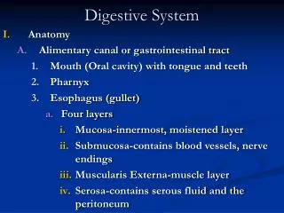
Esophagus • Collapsible • Pierces diaphragm through esophageal hiatus • Contains mucosa, submucosa & muscularis • Superficial layer called adventitia (attaches esophagus to surrounding structures) • Secretes mucous & transports food
Esophagus • Passage of food regulated by esophageal sphincter • Cardiac sphincter relaxes to let food into stomach • Peristalsis (circular muscles contract & longitudinal relax) • Acid reflux is a result of the improper closing of cardiac sphincter
Stomach • J shaped enlargement of the GI tract • Connects esophagus to duodenum • Serves as mixing vat & holding reservoir • Anatomy • 1. Cardia surrounds superior opening • 2. Fundus is superior & left of Cardia • 3. Body (central portion) • 4. Pylorus (connects stomach to duodenum)
Stomach continued • 5. Stomach communicates with small intestine through pyloric sphincter • Has the same basic four layers of the GI tract • Functions • 1. Mixes saliva, food & gastric juice into chyme • 2. Reservoir for holding food • 3. Secretes gastric juice which contains HCL, Pepsin, intrinsic factor & gastric lipase
Stomach continued • 4. HCL kills bacteria & denatures proteins; pepsin begins the digestion of proteins; intrinsic factor aids in absorption of vitamin B12; gastric lipase aids in digestion of triglycerides
Pancreas • Made of clusters of epithelial cells called Acini • Acini secrete pancreatic juice • Composition and function of pancreatic juice • 1. Pancreatic enzymes digest carbohydrates • 2. Trypsin, chymotrypsin, carboxypeptidase and elasatse break down proteins • 3. Pancreatic lipase breaks down triglycerides
Pancreas continued • 4. Ribonucleases & deoxyrobonucleases break down nucleic acids
Liver & Gallbladder • Liver divided into large left & small right lobe by falciform ligament • Liver composed of hepatocytes • Liver has no capillaries but has epithelial lined sinosoids • Sinusoids contain phagocytes (Kupffers cells) • Secretes bile into hepatic duct • Hepatic duct joins cystic duct to form common bile duct
Liver & Gallbladdercontinued • Role & composition off bile • 1. Bile salts (emulsification of lipid) • 2. Biliruben • Functions of liver • 1. Carbohydrate metabolism: maintains a normal blood glucose level by breaking down stored glycogen • 2. Lipid metabolism: Hepatocytes store triglycerides • 3. Protein metabolism: break down amino acids; NH3 converted to urea
Liver & gallbladdercontinued • 4. Synthesis of alpha & beta globulins (antibodies) & fibrogen that helps clot blood • 5. Processing of drugs & hormones: detoxify & chemically alter hormones • 6. Excertion of biliruben: derived from heme portion of broken down blood cells • 7. Synthesis of bile salts: emulsification & absorption of lipids • 8. Storage: glycogen & some vitamins and minerals
Liver & gallbladder continued • 9. Phagocytosis: Kupffers cells phagocytize bacteria & old red and white blood cells
Small intestine • Major sire of digestion & absorption • Begins at pyloric sphincter & ends at large intestine • Anatomy • 1. Duodenum (shortest region) • 2. Jejunum (about 3 ft long & extends to ileum • 3. Ileum (last and largest part of the small intestine; joins large intestine at ileocecal sphincter
Small Intestinecontinued • Histology • 1. Mucosa forms fingerlike villi that increase surface area • 2. Simple columnar epithelium with many microvilli and goblet cells • Functions • 1. Mixes chyme • 2. Peristalsis • 3. Completion of digestion • 4. Absorbs 90% of nutrients
Small intestine continued • Roles of intestinal juices • 1. Mix with pancreatic juice to absorb nutrients from chyme
Chemical digestion in small intestine • Carbohydrates 1. Starches not broken down by amylase are broken down by pancreatic amylase 2. Sucrase breaks sucrose into glucose and fructose 3. Lactase breaks lactose into glucose and galactose 4. Maltase breaks maltose into glucose and glucose
Chemical digestion in small intestine continued • Proteins 1. Starts in stomach by pepsin 2. Trypsin, chemotrypsin, carboxypeptidase and elastase breaks down proteins into peptides 3. Amino peptidase cleaves terminal Amino acids 4. Dipeptidase splits dipeptides into Amino Acids
Chemical digestion in small intestine continued • Lipids 1. Lipases split triglycerides into fatty acids and glycerol 2. Bile salts emulsify triglycerides, which increase surface area exposed to pancreatic lipases 3. Pancreatic lipases and gastric lipases cleave off two fatty acids chains leaving a monoglyceride
Absorption in small intestine • Monosaccharides 1. 120 grams per hour 2. passes through lumen by facilitated diffusion or active transport 3. move out of epithelial cells into capillaries
Absorption in small intestine continued • Amino acids, dipeptides, and tripeptides 1. Absorbed by active transport in duodenum, and jejunum 2. Half absorbed from food and half from dead cells 3. 95-98% absorbed 4. enter blood by diffusion 5. if not filtered and used by hepatocytes they enter general circulation
Absorption in small intestine continued • Lipids 1. By simple diffusion 2. Monoglycerides follow same path as nucleic acids
Large intestine • Anatomy 1. Ileocecal sphincter allows material to pass into large intestine 2. Cecum a. pouch about 2.4 cm long b. Has appendix attached 3. Colon a. Divided into ascending, transverse descending and sigmoid portions
Large intestine continued • Colon continued b. Hepatic flexure (ascending) c. Splenic flexure ( descending) d. Sigmoid enters rectum 4. Rectum a. Anal canal b. Guarded by internal and external sphincter
Large intestine continued • Absorption and feces formation 1. Chyme becomes semi-solid after 3-10 hours in large intestine 2. Feces is composed of water, inorganic salts, dead cells, bacteria, unabsorbed digestive material, and indigestible food parts 3. Water is absorbed in large intestine