Download
1 / 51
520 likes | 656 Views
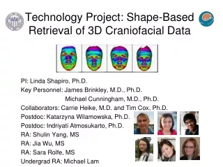
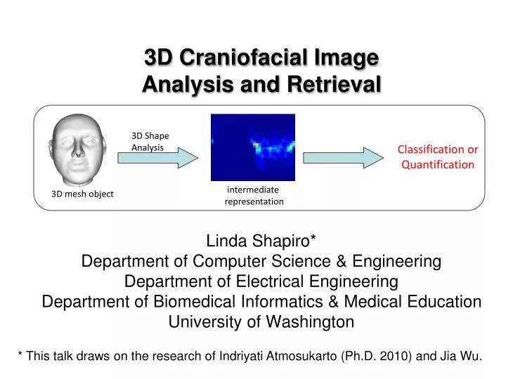
3D Craniofacial Image Analysis and Retrieval. 3D Shape Analysis. Classification or Quantification. intermediate representation. 3D mesh object. Linda Shapiro* Department of Computer Science & Engineering Department of Electrical Engineering

E N D
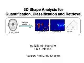
3D Craniofacial Image Analysis and Retrieval 3D Shape Analysis Classification or Quantification intermediate representation 3D mesh object Linda Shapiro* Department of Computer Science & Engineering Department of Electrical Engineering Department of Biomedical Informatics & Medical Education University of Washington * This talk draws on the research of Indriyati Atmosukarto (Ph.D. 2010) and Jia Wu.
Deformational Plagiocephaly • Flattening of head caused by pressure • Delayed neurocognitive development • Assessment is subjective and inconsistent • Need objective and repeatable severity quantification method Plagiocephaly Normal Brachycephaly
22q11.2 Deletion Syndrome (22q11.2DS) • Caused by genetic deletion • Cardiac anomalies, learning disabilities • Multiple subtle physical manifestations • Assessment is subjective
Cleft Lip and Palate • 1:1000 newborns • Wide spectrum of deformities • Varying degrees of symmetry • Objective assessment of severity • and outcome is lacking unilateral cleft lip and palate bilateral cleft lip and palate
Objective • Investigate new methodologies for representing 3D craniofacial shapes • Use these representations for • Classification of abnormality • Quantification of abnormality and particular symptoms • Retrieval of images from a database that are similar to a given image
Deformational Plagiocephaly (Manual) Measurements • Anthropometric landmark • Physical measurements using calipers • Template matching • Landmark photographs Kelly et al. 1999 www.cranialtech.com Cranial Index (CI) Oblique Cranial Length Ratio (OCLR) Hutchison et al. 2005
22q11.2DS (Manual) Measurements • Anthropometric landmark • 2D template landmark + PCA • 3D mean landmark + PCA Boehringer et al. Gabor wavelet + PCA to analyze 10 facial dysmorphologies Hutton et al. Align to average face + PCA
Computing the Plane of Symmetry • Mirror method • Benz et al. 2002 1. hypothesize plane location 2. construct the mirror image 3. overlay mirror image on face 4. find closest corresponding points 5. use corresponding points to better estimate the plane 6. repeat (2-5) till convergence
Data Collection 3dMD multi-camera stereo system Reconstructed 3D mesh
Global 2D Azimuth-Elevation Angle Histogram Indriyati Atmosukarto • 3D Shape Quantification for Deformational Plagiocephaly • Classification of 22q11.2DS • Future retrieval of “similar” normal heads
3D Shape Quantification for Deformational Plagiocephaly • Discretize azimuth elevation angles into 2D histogram • Hypothesis: flat parts on head will create high-valued bins
Shape Severity Scores for Posterior Plagiocephaly • Left Posterior Flatness Score (LPFS) • Right Posterior Flatness Score (RPFS) • Asymmetry Score (AS) = RPFS - LPFS • Absolute Asymmetry Score (AAS)
Classification of Posterior Plagio Absolute Asymmetry Score (AAS) vs Oblique Cranial Length Ratio (OCLR)
Classification of Posterior Plagio Absolute Asymmetry Score (AAS) vs Oblique Cranial Length Ratio (OCLR) #misclassified controls: OCLR 8, AAS 2
Classification of Deformational Plagiocephaly • Treat 2D histogram as feature vector • Classify five plagiocephaly conditions
Classification of 22q11.2DS • Treat 2D histogram as feature vector
Learning 3D Shape Quantification • Analyze 22q11.2DS and 9 associated facial features • Goal: quantify different shape variations in different facial abnormalities
Learning 3D Shape Quantification -Facial Region Selection • Focus on 3 facial areas • Midface, nose, mouth • Regions selected manually
Learning 3D Shape Quantification -2D Histogram Azimuth Elevation • Using azimuth elevation angles of surface normal vectors of points in selected region
Learning 3D Shape Quantification -Feature Selection • Determine most discriminative bins • Use Adaboost learning • Obtain positional information of important region on face
Learning 3D Shape Quantification -Feature Combination • Use Genetic Programming (GP) to evolve mathematical expression • Start with random population • Individuals are evaluated with fitness measure • Best individuals reproduce to form new population
x y 5 + * 5*(x+y) Learning 3D Shape Quantification -Genetic Programming • Individual: • Tree structure • Terminals e.g variables eg. 3, 5, x, y, … • Function set e.g +, -, *, … • Fitness measure e.g sum of square …
Learning 3D Shape Quantification - Feature Combination • 22q11.2DS dataset • Assessed by craniofacial experts • Groundtruth is union of expert scores • Goal: classify individual according to given facial abnormality
Learning 3D Shape Quantification -Feature Combination • Individual • Terminal: selected histogram bins • Function set: +,-,*,min,max,sqrt,log,2x,5x,10x • Fitness measure: F1-measure X6 + X7 + (max(X7,X6)-sin(X8) + (X6+X6))
Learning 3D Shape Quantification - Experiment 1 • Objective: investigate function sets • Combo1 = {+,-,*,min,max} • Combo2 = {+,-,*,min,max,sqrt,log2,log10} • Combo3 = {+,-,*,min,max, 2x,5x,10x,20x,50x,100x} • Combo4 = {+,-,*,min,max,sqrt,log2,log10, 2x,5x,10x,20x,50x,100x}
Learning 3D Shape Quantification - Experiment 1 • Best F-measure out of 10 runs
Tree structure for quantifying midface hypoplasia ((X7-X7) + (X6+(((X6+X6)-X7)+(X7-X2)))+X7))+(X9-5X9+X7+X7) Xi are the selected histogram bins
Learning 3D Shape Quantification - Experiment 2 • Objective: compare local facial shape descriptors
Learning 3D Shape Quantification - Experiment 3 • Objective: predict 22q11.2DS
Learning to Compute the Plane of Symmetry for Human Faces Jia Wu • Overview • Train a classifier to identify regions about landmark points • Train a second classifier to determine which of these • regions are useful in computing the plane of symmetry • Use these classifiers to select regions of a face and • use their center points to compute the plane of symmetry • The RANSAC algorithm fits the plane and discards • outliers
Landmark by medical experts Landmarks labeled by experts Standard symmetry plane
10 kinds of landmarks. • Nose: ac, prn, sn,se • Eyes: en, ex • Mouth: (li,ls), ch, sto, slab
Positive/negative samples Training for en: the inner corners of the eyes Training for prn: most protruded point of nasal tip
Features: Histograms of Gaussian Curvature andCurvedness with Different Neighborhood Sizes Gaussian curvature Gaussian curvature Gaussian curvature Gaussian curvature Gaussian curvature Gaussian curvature Gaussian curvature Curvedness (distance from origin in curvature plane) Histograms of Gaussian curvatures of a positive sample and a negative sample
Interesting points prediction Prediction of en: the inner corners of the eyes Prediction of prn: most protruded point of nasal tip
Connected regions Connected regions for en: each color means one region Connected regions for prn: each color means one region
How to define “useful” symmetric regions • A useful pair of regions should be symmetric to the standard symmetry plane • A useful single region should have the center on the standard symmetry plane useful regions for en useful regions for prn
Procedure for New data Select possible landmark areas( from Landmark model) Find and pair connected regions interesting regions for prn Determine useful singles and useful pairs ( from Symmetry model) Get center and draw a plane using learned centers Predicted as useful single Predicted as useful pair
Procedure for New Images Centers of useful regions Centers for constructing plane of symmetry are red. Result: Plane of symmetry
Results on 22q11.2DS DataSet compared to Mirror Method worst best worst best
Results on Rotated Data Set worst best worst best
Some Results on Cleft Subjects Learning method Mirror method
Contributions • Representation of craniofacial anatomy by azimuth elevation histograms and other local features. • Classification and quantification of abnormal conditions using this representation. • New learning methodology for finding the plane of symmetry of the face, even for cleft patients.
Future Directions • A retrieval system is being designed that will use multiple different low-level features in different areas of the face to retrieve similar images from image databases. • The asymmetry of the face will be studied more thoroughly and quantified. • A series of new features will be designed to describe and quantify the degree of cleft lip and palate.
Acknowledgements This research was supported by • the National Science Foundation under grant number DBI-0543631 • the National Institute of Dental and Craniofacial Research under grant number 1U01DE020050 as part of the FaceBase Consortium • the National Institute of Health under grant number K23-DE017741