Download
1 / 46
470 likes | 548 Views
Hemostasis. Hemostasis depends on the integrity of Blood vessels Platelets Coagulation factors Anticoagulation factors. Initial Laboratory Tests For Bleeding Abnormalities. Platelet count. Bleeding time. Partial thromboplastin time (PTT). Prothrombin time (PT). Thrombin time(TT).

E N D
Hemostasis • Hemostasis depends on the integrity of • Blood vessels • Platelets • Coagulation factors • Anticoagulation factors
Initial Laboratory Tests For Bleeding Abnormalities • Platelet count. • Bleeding time. • Partial thromboplastin time (PTT). • Prothrombin time (PT). • Thrombin time(TT).
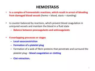
Causes of Abnormal Bleeding • Vascular disorders. • Plateletcauses: (Thrombocytopenia, functional defects). • Defective coagulation.
Vascular Bleeding Disorders • Easy bruisability and bleeding from small vessels. • Caused by abnormal vessels or abnormal perivascular connective tissue. • Sites of bleeding: skin and mucous membranes. • Bleeding time and other screening tests are normal. • Congenital or acquired.
Senile purpura due to atrophy of perivascular connective tissue. Scurvy. Steroid purpura (defective CT). Hereditary hemorrhagic telangiectasia Ehlers-Danlos syndrome. Vasculitis: -Hypersensitivity: Henoch-Schoenlein purpura - Septic: infections. measles dengue meningococcemia ricketsial infections Acquired Vascular Defects
Platelet causes • Thrombocytopenia (quantitative) • Platelet function disorders (qualitative)
Relationship Between Platelet Count and Bleeding • Normal range 150-400X103 per µl. • Levels above 60x103/µl will not cause bleeding under normal conditions. • Levels below 20x103/µl will cause: Petechiae, mucosal bleeding. Post-operative bleeding, CNS bleeding. • Levels around 5x10³/µl can lead to fatal CNS or GI hemorrhage. • Levels between 20 and 60x103/µl may cause bleeding (depending on platelets functional status).
Classification of Thrombocytopenia • Failure of production • Increased platelet destruction • Sequestration: hypersplenism • Dilutional
Decreased BM megakaryocytes Infiltrative diseases of BM Aplastic BM Amegakaryocytic thrombocytopenia HIV infection Drugs Increased BM megakaryocytes Megaloblastic anemia Myelodysplasia Alcohol induced Rare diseases such as Wiskott-Aldrich syndrome Thrombocytopenia Due to Failure of Production
Thrombocytopenia Due to Increased Platelets Destruction • ITP • Secondary immune thrombocytopenia as in SLE • Drug related immune thrombocytopenia as in quinidine and heparin. • Post transfusion thrombocytopenia • Neonatal thrombocytopenia either due to autoantibodies or alloantibodies • DIC and microangiopathic hemolytic anemia
Chronic Immune Thrombocytopenic Purpura (ITP) • High incidence in women of child bearing age. • Mostly idiopathic, secondary causes include SLE, HIV, CLL, Hodgkin’s disease. • Autoantibodies against GP IIb/IIIa, or Ib/IX. • Platelets lifespan reduced to hours. • Megakaryocytes increased. • Petechial bleeding, easy bruising, menorrhagia.
ITPDiagnosis • Decreased platelet count (10-50x109/l). • Hb. and WBCs are normal. • Increased megakaryocytes numbers in BM. • Antiplatelet antibodies. • ? Antinuclear antibodies in SLE. • ? Coomb’s test in Evan’s syndrome.
ITPTreatment • Steroids. • Splenectomy. • High dose IV immunoglobulins. • Immunosuppressive therapy. • Platelets transfusion: Not helpful
Acute ITP • Affects children. • Preceded by infection or vaccination in 75% of cases. • Spontaneous remission in >90% of cases. • Severe cases benefit from steroids or IV immunoglobulins.
Thrombotic Microangiopathies • Widespread formation of thrombi in the microcirculation composed of platelets and fibrin. • Consumptive thrombocytopenia and microangiopathic hemolytic anemia • TTP: 5 criteria • HUS; usually no CNS caused by endothelial injury secondary to E.coli O157:H7 infection in children, elderly
TTP • Deficiency of metalloprotease (ADAMTS13) needed for cleaving very hmw vWF (usually acquired autoAb, but may be inherited) • Accumulation of vhmw vWF leading to thombi • Thrombocytopenia, fever, CNS, renal microangiopathic hemolytic anemia. • Normal PT, PTT • Plasma exchange.
Platelet Membrane Glycoproteins • GP Ia-IIa: adhesion to collagen. • GP Ic-IIa: laminin receptor. • GP IIb-IIIa: binding to fibrinogen. • GP Ib-IX: adhesion to subendothelial tissue via vWF.
Qualitative disorders of platelets (1) Bernard Soulier syndrome • Adhesion Defect • Autosomal recessive. • Deficiency of GpIb/IX (CD42) which serves as the receptor for vWF. • Giant platelets.
Qualitative disorders of platelets 2 • Glanzman’s thrombasthenia • Aggregation defect • Failure to aggregate in response to ADP, collagen, epinephrine and thrombin • Autosomal recessive • Deficiency of GpIIb/IIIa (CD41/CD61) which serves as the receptor for fibrinogen. • Storage pool diseases • Initial aggregation normal, but secretion of thromboxane, ADP, and other granules contents impaired
Coagulation disorders • Congenital or acquired • Acquired: are the most common many factors simultaneously deficient -- Vit. K deficiency: II, VII, IX, X -- Liver disease (most factors synthesized in the liver)
von Willebrand Factor (vWF) • Coded by a gene located on the short arm of chromosome 12 • The primary ptn. product is a 250,000 Da dimer. • Transformed by multiple disulfide bonds to form a series of multimers ranging in weight from 10,000,000 to 20,000,000 Da
vWF • Adhesive protein, bridges collagen to platelets receptor GPIb • Carrier protein to factor VIII • Ristocetin induces platelets agglutination in the presence of vWF • Stored in Weibel-Palade bodies of endothelial cells, and a granules of platelets
von Willebrand Disease • The most common hereditary bleeding disorder (prevalence 0.1-1%). • Mild bleeding problems • Mucous membrane bleeding • Easy bruising • menorrhagia • Post-operative bleeding • Most cases are inherited as autosomal dominant disorder. • Prolonged bleeding time
von Willebrand Disease type I • The most common type (75% VWD cases). • Autosomal dominant disorder with variable penetrance (60%) • Reduced circulating vWF and factor VIII. • All sizes of vWF multimers are present • Slight prolongation of APTT • Platelets fail to agglutinate by ristocetin
von Willebrand Disease type II • Reduced circulating large and intermediate multimers of vWF. • vWF hmw is missing in type A • vWF has hightened interaction with GP1b in type B • Fctor VIII level is normal. • Failure of agglutination by ristocetin in IIa. • smaller ristocetin doses cause aggregation of platelets in type IIb. • Platelet type VWD is similar to type IIb, but GPIb is defective. Cryo precipitate alone can induce agglutination of platelets
von Willebrand Disease type III • Absent plasma vWF • Markedly reduced factor VIII coagulant activity • Autosomal recessive inheritance
von Willebrand Disease type IIN • Formerly known as VWD Normandy • Mutation in vWF affecting association with factor VIII • Manifestations similar to hemophilia, but transmission is autosomal recessive • Should be suspected in hemophilia patients if: • Inheritance is not sex-linked • Poor response to factor VIII infusion
von Willebrand Disease Manegement • DDAVP which releases vWF from endothelial cells. • Single donor plasma concentrate. • Factor VIII concentrate or cryoprecepitate for type III von Willebrand disease
Inherited Deficiencies of Coagulation Factors Deficiency Fibrinogen Prothrombin Factor V Factor VII Factor VIII Factor IX Factor X Factor XI Factor XIII Incidence in Population 1:1 million 1:2 million 1:1 million 1:500,000 1:10,000 1:60,000 1:1 million 1:1 million 1:2 million Gene on Chromosome 4 11 1 13 X X 13 4 6 & 1 Mode of Inheritance AR AR AR AR XLR XLR AR AR AR
Hemophilia A • Incidence: one in 10,000. • Sex-linked. 1/3 of the cases have no family history (recent mutation or generations of silent carriers). • Caused by absolute reduction of factor VIII or normal amount but defective factor VIII. • Severity of disease depends on factor VIII level • Normal level 100 U/dl • Severe cases level <2 U/dl • Moderate cases level 2-5 U/dl • Mild cases level 5-25 U/dl
Hemophilia A • Sites of bleeding: • Large joints and soft tissue • Urinary tract and GI tract • Brain • Nose • Laboratory tests: • Prolonged PTT. Normal PT and TT. • Low factor VIII assay. • Treatment: replacement therapy (factor VIII concentrate or recombinant VIII). • Inhibitor antibodies develop in 18-50% of patients
Nomenclature of Factor VIII Protein lacking or aberrant in hemophilia A Functional property of factor VIII missing in hemophilia A, measured by coagulation assays Antigenic property of factor VIII, measured by immunoassays Factor VIII Factor VIIIc Factor VIIIAg
Factor VIII (FVIII) and the FVIII Gene • The gene is located at the tip of the X chromosome. • Huge gene composed of 26 exons. • FVIII mRNA is ~9kb, and encodes a 300kD protein. • FVIII is acofactor for F IX in the proteolytic activation of F X. • Bound to vWF in plasma. • Markedly unstable in the absence of vWF.
Molecular Genetics of Hemophilia A • Point mutation in exons or splice junctions (~50%) • Inversion of intron 22 (~45%) • Deletions or less commonly insertions (~5%)
Hemophilia in Females Exceedingly rare, seen in: • Mating between a carrier mother and affected father • Carriers with abnormalities of X-chromosome • Extreme lyonization • X mosaicism or deletion • Newly mutant gene
Hemophilia B • Incidence: one in 60,000. • Sex-linked. • Severity of disease depends on factor IX level • Normal level 100 U/dl • Severe cases level <2 U/dl • Moderate cases level 2-5 U/dl • Mild cases level 5-25 U/dl • Bleeding sites: similar to hemophilia A. • Laboratory tests: • Prolonged PTT. Normal PT and TT. • normal factor VIII assay.
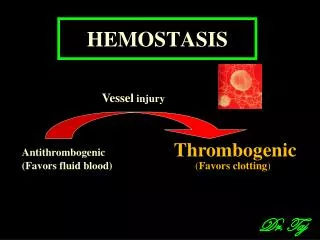
Disseminated Intravascular Coagulation (DIC) • Widespread thrombi in the microcirculation with secondary consumption of platelets and coagulation factors (consumptive coagulopathy) • acute, subacute and chronic • Relase of tissue factor or endothelial damage: activation of extrinsic coag. cascade and dec. inhibitory pathways
DIC (Consumptive Coagulopathy) • Thrombosis, fibrinolysis, bleeding. • Obstetric complications; infections; cancers (panc, lung, prost, stomach, APML), massive tissue injury (trauma, burn), others (shock, liver, heat stroke, vasculitis, intravascular hemolysis). • Lab: Low plat and fibrinogen, high PT. PTT and FDP
DIC • Microangiopathic hemolytic anemia, infarcts, hemorrhages • microthrombi and infarcts in most organs esp. kidneys, brain, adrenals, heart • Small infarcts in kidneys, but in severe cases cortical necrosis • Adrenal, pituitary, placenta • Clinical: bleeding in acute, thrombosis in chronic; minimal, or severe shock, coma