Download
1 / 86
1k likes | 1.47k Views
B CELL DEVELOPMENT AND ACTIVATION. In healthy people, there are mature B cells with the capacity to make antibodies to virtually any antigen. Bone marrow is the primary lymphoid organ in which B cell development occurs. .

E N D
B CELL DEVELOPMENT AND ACTIVATION • In healthy people, there are mature B cells with the capacity to make antibodies to virtually any antigen. • Bone marrow is the primary lymphoid organ in which B cell development occurs.
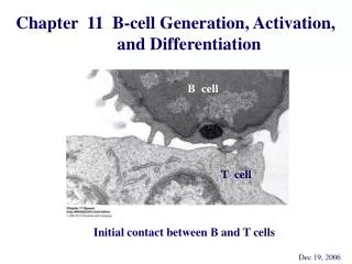
Bone marrow is the primary lymphoid organ in which B cell development occurs. • Following initial development in bone marrow, mature B cells migrate to various secondary lymphoid tissues, including lymph nodes, spleen, gut-associated lymphoid tissue and blood. • There, mature B cells can interact with antigen, become activated, and further differentiate into antibody-secreting cells
B and T cells undergo distinct differentiation pathways. • B cells are generated in the bone marrow, with mature B cells, which are ready to respond to antigen, then exiting and migrating to lymph nodes and spleen. T cells are generated in the thymus.
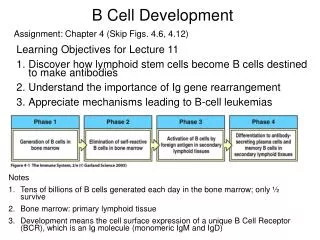
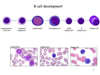
The development of B cells, starting from hematopoietic stem cells and ending with cells that produce antibodies, can be divided into four phases:
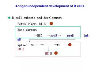
Phase 1 – development of B cells in bone marrow • This first phase of B cell development is the generation of B cells in bone marrow. • There, stem cells develop into pro-B cells, then pre-B cells, and finally mature B cells, which exit the bone marrow and migrate to secondary lymphoid organs. • This phase of B cell development is not driven by contact with antigen: antigen independent. • The DNA rearrangements that result in a functional cell-surface immunoglobulin molecule occur during this phase.
Stem cells have both their H-chain and L-chain genes in germ-line, un-rearranged configuration • The earliest cell that has made the commitment to the B cell lineage is the pro-B cell: pro-B cells have begun to rearrange their H-chain gene.
Once a B lineage cell expresses cell surface H-chain (m) it is defined as a pre-B cell. • However, the early pre-B cell receptor is not the final form of surface immunoglobulin: H-chain + surrogate light chain (molecule that mimics L-chain)
Further DNA rearrangements result in the formation of a functional L-chain, then IgM (H-chain + L-chain) is expressed on the cell surface. • When a cell has productive rearrangements of both H- and L-chains) it becomes an immature B cell:
This first phase of B cell development in bone marrow is dependent on association with stromal cells. • Stromal cells are non-lymphoid cells that provide an appropriate microenvironment for B cell development. • Bone marrow stromal cells produce both cell surface-stimulatory molecules, as well as growth factors and cytokines, which help drive B cell development.
For a B cell to survive this phase of development, it must have productive rearrangements of both H-chain and L-chain. • Failure to do this results in cell death - cells that have unproductive rearrangements (such as rearrangements that are not in a correct reading frame) are eliminated. • A given B cell can undergo repeated rearrangements.
The rearrangements that result in functional H- and L-chains occur in a specific order:
Expression of a functional B cell receptor protein on the cell surface stops further rearrangement of the gene encoding that product:
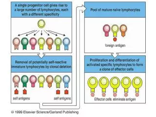
Phase 2 – elimination of self-reactive cells • Once B cells express a functional cell surface receptor for antigen (immature/mature B cell stage) they have the potential to be stimulated by contact with antigen, and to become antibody-secreting cells. • Since the DNA rearrangements that result in functional H-chain and L-chain are not antigen-driven, a fraction of immature B cells will have a BCR that, by chance, reacts with some component of self - self antigen reactive cells • These immature B cells are removed by clonal deletion, either in the bone marrow, or shortly after leaving the bone marrow.
Encounter with self-antigen results in apoptosis (death), or anergy (unresponsiveness) • B cells that survive this step express surface IgD as well as IgM: mature B cells Goodnow experiments
After elimination of self-reactive B cells, mature cells, which express both cell surface IgM and IgD, are ready to leave the bone marrow, can interact with antigen in secondary lymphoid organs.
Phase 2 – elimination of self-reactive B cells, generation of mature IgM+, IgD+ cells:
Phase 3 – activation of B cells on contact with antigen • Following the generation of a functional B cell receptor for Ag, and the removal of self-reactive cells, mature Ag-responsive B cells(IgM+, IgD+) emigrate from bone marrow. • These mature B cells go to secondary lymphoid organs.
In the lymph node, B cells gather in primary lymphoid follicles, where they receive viability-promoting signals, interacting with follicular dendritic cells, and wait for antigen • B cells enter lymph nodes via high endothelial venules (HEV) to reach these primary follicles. • B cells can recirculate out via the lymphatic circulation, and back into blood.
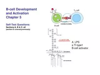
What is meant by ‘B cell activation’? • Go -> G1 of cell cycle (increase in size) • upregulate • MHC class II • costimulatory molecules (B7-2) • adhesion molecules (ICAM1) • cytokine receptors (IL-2R) • migrate to outer T zone • altered response to chemokines • become receptive to T cell help • protected from fas • enter mitosis if provided with submitogenic doses of other stimuli (LPS, CD40L, IL-4)
Types of antigen • T-independent (TI) antigens - Type I • induce division/differentiation independently of BCR (polyclonal mitogens) • LPS, bacterial (CpG) DNA • T-independent (TI) antigens - Type II • induce division/differentiation by BCR signaling alone • bacterial polysaccharides, repeating surface molecules on viruses • T-dependent (TD) antigens • activate via BCR but depend on additional signals from helper T cells to cause division/differentiation • any antigen containing protein • Most pathogens contain both T-I and T-D antigens • Only TD antigens can induce Germinal Center responses
Types of B cell Antigens T-independent (TI) T-cell dependent (TD) T cell present Ag mitogenic 'activation' signal mitogenesis BcR signal but not mitogenic differentiation -> most pathogens contain both T-independent and T-dependent antigens
Innate features of pathogens act as B cell costimulators • pathogen multivalency • provides a level of BCR crosslinking optimal for activation • many pathogens activate TLRs • TLR signaling synergizes with BCR signal • many pathogens activate the complement cascade and become C3d coated • complement receptor (CR) crosslinking synergizes with BCR signal
Antigen-C3d complexes cross-link BCR and CR2-CD19 complex - increase sensitivity to antigen
T-Independent (type II) Responses Emerging Model: DC DC plasma cells B cell B cell Current Paradigm : Multivalent Antigen B cell B cell plasma cells
T-Dependent Responses Antigen DC T cell T cell B cell plasma cells B cell binds Ag via surface Ig, transmits BCR signals and presents peptides to T cells, receives T cell help (growth and differentiation factors) Secretes Antibody (Ab) Dendritic Cell (DC) internalizes antigen (Ag), processes into peptides, presents peptides together with MHC molecules to T cells
Antigen-specific B cells are detained in the T cell-areas, where they interact with antigen, and with antigen-specific activated helper T cells. Stimulated antigen-specific B cells then proliferate and differentiate, eventually forming plasma cells and germinal centers:
B cells are antigen-presenting cells • BCR cross-linking induces antigen internalization to endosomes • antigen is proteolysed to peptides • peptides associate with MHC class II • MHC class II-peptide complexes traffic to surface of B cell\ • B cells present antigen recognized by their BCR ~105 x more efficiently than other antigens
B cell antigen presentation and the concept of linked help protein sugar T Protein Specific T cell Sugar Specific B cell Antigen internalization, proteolysis -> presentation of peptides
Interactions between antigen-specific B and T cells1 day after HEL antigen injection HEL-specific (Ig-tg) B cells HEL-specific (TCR7 tg) T cells
Onset of B-T interaction HEL-specific B cells HEL-specific T cells
B cells can interact with multiple T cells HEL-specific B cells HEL-specific T cells
However, signaling via the BCR alone is not sufficient to activate the B cell - • Second signals (co-stimulatory signals) are necessary for activation.
-or-deficient mice and human do not undergo isotype switching They only have IgM: hyper IgM syndrom
Phase 4 – differentiation to antibody-secreting cells • Some of the progeny of these antigen-activated B cells differentiate into IgM-secreting plasma cells (antibody-secreting cells).
Plasma cells: • terminally-differentiated cells • derived from activated B cells or memory cells • loaded with endoplasmic reticulum • devoted to protein (antibody) synthesis • no longer express surface immunoglobulin or MHC class II • no longer responsive to antigen contact • live for several weeks • migrate away from the site of initial contact with helper T cells, either to the medullary cords of the lymph nodes or to the bone marrow
B Mature B cell B Plasma cell Plasma cells Surface Surface High rate Growth Somatic Isotype Ig MHC II Ig secretion hypermut’n switch High YesNo Yes Yes Yes Low No Yes No NoNo
Other antigen activated B cells give rise to germinal centers (GC), zones of proliferating activated B cells:
These germinal centers (GC) contains: • proliferating (D - centroblasts) B cells express low levels of Ig, especially IgD • differentiating (L - centrocytes) B cells express high levels of Ig
B cells can interact with an antigen that is bound to the surface of follicular dendritic cells in the lymph nodes. • These cells trap and concentrate antigen, maximizing the interaction of antigen with B cells
Follicular Dendritic cells (stained blue)in the Germinal Centre
Retention of Antigens on Follicular Dendritic Cells Radiolabelled antigen localises on the surface of Follicular Dendritic cellsand persists there, without internalisation, for very long periods
Club-shaped tips of developing dendrites Filiform dendrites Bead formation on dendrites Bead formation on dendrites Maturation of Follicular Dendritic cells
DC veils Iccosomes (black coated particles) bind to and are taken up by B cell surface immunoglobulin Iccosome formation and release The veils of antigen-bearing dendritic cell surround the beads and the layer of immune complexes is thickened by transfer from the dendritic cell. These beads are then released and are then called ICCOSOMES
Anti- B cell Y Y B Y Uptake of Iccosomes/Antigen by B cells Iccosomes bearing different antigens CD40 Surface Ig captures antigen Cross-linking of antigen receptor activates B cell Activated B cell expresses CD40
B B Fate of Antigens Internalised by B cells 1. Capture by antigen specific Ig maximises uptake of a single antigen 2. Binding and internalisation via cell surface Ig induces the expression of CD40 3. Antigen enters exogenous antigen processing pathway and is degraded 4. Peptide fragments of antigen are loaded onto MHC molecules intracellularly. MHC/peptide complexes are then expressed at the cell surface
This selection process involves competition for both antigen and for helper T cells. • Antigen is trapped on the surface of follicular dendritic cells in the form of immune complexes (antigen + antibody complexes). • B cells that bind to Ag with high affinity live, others die by apoptosis.
Centrocytes interact with T cells by presenting processed antigen to them via their MHC class II molecules. • Centrocytes that receive co-signaling (via CD40, MHC class II and cytokines), as well as signaling via their antigen receptor survive • Centrocytes that do not bind antigen and T cells with sufficient affinity die by apoptosis.