Download
1 / 37
370 likes | 760 Views
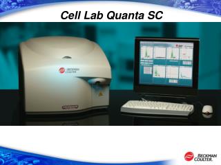
Cell Lab Quanta SC. Cell Lab Quanta - Technology. Technology – Coulter Volume. The Coulter Counter measures the electrical resistance across an aperture immersed in an electrolytic buffer. When a cell passes through the aperture, the resistance increases

E N D
Technology – Coulter Volume The Coulter Counter measures the electrical resistance across an aperture immersed in an electrolytic buffer. When a cell passes through the aperture, the resistance increases and an “event” is recorded as an electrical potential pulse. Coulter volume measurement remains one of the most accurate ways to measure cell size.
Aperture Current Electrolytic Solution Electrodes + _ Technology – Coulter Volume Example
Technology – Patented Flow Cell Natural shear forces create 2µ flow stream 125μm 125μm Technology – Flow Cell Stream stability yields <30% increased “optical” resolution and “electrical Volume” sensitivity
Technology – flow cell Electrodes on both sides of the sample chamber establish an electric current, which when disrupted by cells both count and size the population Epi-illumination identical to principle of inverted microscopy systems carries excitation and emission light for UV Light and emission of 488 excited light
Technology – Versatile arc lamp excitation source The four primary excitation wavelengths from the arc provide a flexible solution to encompass many common excitable dyes, including UV.
Technology- Filter Block 488 Standard Applications UV Arc Lamp Setup
Technology- Syringe Mechanism • Features • Accurate delivery of sample to flow cell • User Controlled Sample Injection Rate • 4.17 to 100 ul per minute • Benefits • Automatic, Accurate Enumeration of Sample • Do not need to run Beads with sample • Minimal sample used • Unused sample can be recovered
Diameter vs. Volume Increase in Diameter 5% 25% 16% 100% Increase in Volume This is the reason why volume analysis affords significant sensitivity over typical forward light scattering.
ECV Light Scatter Light Scatter vs. Volume
FC= Fluorescence/Volume of Cell Fluorescence Concentration Control Bref A Treated Normal Fluorescence Readout Fluorescence normalized for Cell Volume Note: Assay conditions unoptimized.
Cell Viability Apoptosis: Annexin-5, Caspase 3 Cell Cycle: UV, Cyclin A2, PhosphoHistone Stem Cell Side Population Analysis Three Color Immunophenotyping Bacteria and other submicron populations Gene Expression: GFP, YFP, CFP transfections Spermatozoa Viability Identification of marine organisms Plant Ploidy Analysis And many more….. Applications for the Cell Lab Quanta
Viability and Electronic Volume Total Distribution of Cells by Electronic Volume
Size Distribution Cells Gated by Viable vs Non Viable Viable Non-Viable
Viability – 7AAD Untreated 94% Viable Anti CD95 Treated 75% Viable
Side Scatter vs Volume Clear Separation Side Scatter Electronic Volume
Annexin V+ PI+ (late apoptotic/ necrotic) Annexin V+ PI- “early apoptotic” “viable” cells Apoptosis Annexin-7AAD Control Jurkat Cells Treated cells (induced) Thapsagarin
Apoptosis- Active Caspase 3 Control Anti-CD95 treated Jurkat cells
Cell Cycle Cyclin D + CDK2/4 RB Phosphorylation Cyclin E+ CDK2 RB Phosphorylation Cyclin A + CDK2 Cyclin B/A + CDC2
The Cell Lab Quanta is the highest resolution system on the market for cell cycle analysis. Obtain G0/G1, S-phase, G2+M statistics plus cell or nuclear volume (size). Applications: UV Cell Cycle • DNA dyes used on the Quanta Include: • DAPI • Propidium Iodide • Draq5 • Hoechst • 7-AAD Region Name Diameter MCV Counts Pct FL1 Mean FL1 HPCV CV1 Color 125.7 G0/G1 6.22 10,901 62,21% 191.4 2.78 4.86 S-Phase 7.06 183.9 4,313 24.61% 265.3 6.07 11.71 G2 7.58 228.4 1,664 9.50% 351.1 4.30 3.79
Drug Effect on Cell Cycle Control Colchicine Camptothecin Anti CD95
Two Color Cell Cycle- 488 Laser Cyclin A2 FITC DNA- 7-AAD Control Treated
Two Color Cell Cycle- 488 Laser M Phase 2% M Phase 19% Phospho-Histone H3 DNA- 7-AAD Control Treated
Stem Cell Analysis (Side Population) Blue Hoechst 33342 Red Hoechst 33342 Applications: Stem Cell Research Hematopoietic stem cells (HSC) are multipotent cells that reside in the bone marrow and replenish all adult hematopoietic lineages throughout the lifetime of the animal. Studies in the hematopoetic and muscle systems show that stem cells have the ability to efflux the dye Hoechst 33342. Cells with this phenotype are referred to as the side population (SP). In many cases, this population represents less than 1 % of the total population. (Goodell et al. (1996)) Historically this analysis was done on very expensive sorter systems due to requirement of Hoechst 33342 UV excitation. This very same analysis can be done on the Cell Lab Quanta at a fraction of the cost and with the ability to give additional statistics not provided on traditional cytometry systems. Counts Region Name Diameter MCV [ ] 105 Pct Color All Cells 7.2 194.5 9.51 97.46% 97,459 SP Cells 7 182.9 639 6 0.64% Other NA NA 1,902 NA 1.90%
Three Color Immunophenotyping Lymphocytes stained with CD4-FITC, CD8-PE and CD3-PC5
BacLight Green (Syto 9) stains live and dead cells Permeable dye that stains nucleic acids Dye is excited with the 488nm laser Used to trigger for enumeration Bacteria Staining
Bacteria Viability- E. Coli PI Volume Syto 9 PI- Dead Syto9- Live Syto 9/PI
Live/Dead Enumeration 2.8% 50.4% Live Live with Dead Cells
Budding Yeast Cells- EV, SS, FL1 Stained with Syto 9 and triggered on FL1
Dynamic range for volume measurement 125 Micron Flow Cell: 3-40 micron diameter particles 50 Micron Flow Cell: 0.78.-6 (In progress) Dynamic range for counting 2x10⁶ to 3x10⁴ Light Source 488nm Laser 2-22mW Mercury Arc Lamp 365, 405, and 435 lines Detection Three PMTs for detection from 400-800nm Filters for Blue, Green , Orange, and Red Fluorescence 465/30, 525/30, 575/30, 670 Long Pass Photodiode for Side Scatter Detection Key Specifications