Download
1 / 148
1.48k likes | 1.49k Views
3. Cells and Tissues. Concepts of the Cell Theory (no notes). A cell is the basic structural and functional unit of living organisms. The activity of an organism depends on the collective activities of its cells.

E N D
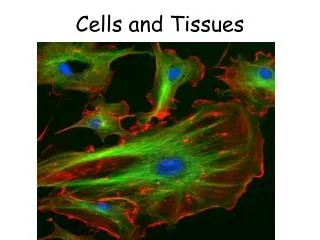
3 Cells and Tissues
Concepts of the Cell Theory (no notes) A cell is the basic structural and functional unit of living organisms. The activity of an organism depends on the collective activities of its cells. According to the principle of complementarity, the biochemical activities of cells are dictated by the relative number of their specific subcellular structures. Continuity of life has a cellular basis.
Chemical Components of Cells (no notes) Most cells are composed of the following four elements Carbon Hydrogen Oxygen Nitrogen
Cells and Tissues 1. Carry out all chemical activities needed to sustain life 2. Cells are the building blocks of all living things. 3. Tissues are groups of cells that are similar in structure and function.
Anatomy of the Cell 1. Cells are not all the same. 2. All cells share general structures. 3. All cells have three main regions Nucleus Cytoplasm Plasma membrane
Nucleus Cytoplasm Plasma membrane (a) Figure 3.1a
The Nucleus 1. Control center of the cell Contains genetic material (DNA) 2. Three regions Nuclear envelope (membrane) Nucleolus Chromatin
Nuclear envelope Chromatin Nucleus Nucleolus Nuclear pores Rough ER (b) Figure 3.1b
The Nucleus Nuclear envelope (membrane) 1. Barrier of the nucleus 2. Consists of a double bilayer membrane 3. Contains nuclear pores that allow for exchange of material with the rest of the cell
The Nucleus Nucleoli 1. Nucleus contains one or more nucleoli and are the site of ribosome assembly 2. Ribosomes migrate into the cytoplasm through nuclear pores
Plasma Membrane 1. Barrier for cell contents 2. Double phospholipid layer Hydrophilic heads (water-loving) Hydrophobic tails (water-hating) 3. Also contains proteins, cholesterol, and glycoproteins
Extracellular fluid (watery environment) Glycoprotein Glycolipid Cholesterol Sugar group Polar heads of phospholipid molecules Bimolecular lipid layer containingproteins Channel Nonpolar tailsof phospholipid molecules Proteins Filaments of cytoskeleton Cytoplasm (watery environment) Figure 3.2
Microvilli Tight (impermeable) junction Desmosome (anchoring junction) Plasma membranes of adjacent cells Connexon Gap (communicating) junction Underlying basement membrane Extracellular space between cells Figure 3.3
Cytoplasm 1.Contains three major elements A. Cytosol Fluid that suspends other elements B. Organelles Metabolic machinery of the cell “Little organs” that perform functions for the cell C. Inclusions Chemical substances such as stored nutrients or cell products
Chromatin Nuclear envelope Nucleolus Nucleus Plasma membrane Smooth endoplasmic reticulum Cytosol Lysosome Mitochondrion Rough endoplasmic reticulum Centrioles Ribosomes Golgi apparatus Secretion being released from cell by exocytosis Microtubule Peroxisome Intermediate filaments Figure 3.4
Cytoplasmic Organelles Mitochondria “Powerhouses” of the cell Change shape continuously Carry out reactions where oxygen is used to break down food Provides ATP for cellular energy
Cytoplasmic Organelles Ribosomes Made of protein and RNA Sites of protein synthesis Found at two locations Free in the cytoplasm As part of the rough endoplasmic reticulum
Cytoplasmic Organelles Endoplasmic reticulum (ER) Fluid-filled tubules for carrying substances Two types of ER Rough endoplasmic reticulum Studded with ribosomes Synthesizes proteins Smooth endoplasmic reticulum Functions in lipid metabolism and detoxification of drugs and pesticides
Ribosome mRNA 1 As the protein is synthesized on the ribosome, it migrates into the rough ER cistern. Rough ER In the cistern, the protein folds into its functional shape. Short sugar chains may be attached to the protein (forming a glycoprotein). 2 1 3 2 Protein The protein is packaged in a tiny membranous sac called a transport vesicle. 3 4 Transport vesicle buds off The transport vesicle buds from the rough ER and travels to the Golgi apparatus for further processing. 4 Protein inside transport vesicle Figure 3.5
Ribosome mRNA 1 As the protein is synthesized on the ribosome, it migrates into the rough ER cistern. Rough ER 1 Protein Figure 3.5, step 1
Ribosome mRNA 1 As the protein is synthesized on the ribosome, it migrates into the rough ER cistern. Rough ER In the cistern, the protein folds into its functional shape. Short sugar chains may be attached to the protein (forming a glycoprotein). 2 1 2 Protein Figure 3.5, step 2
Ribosome mRNA 1 As the protein is synthesized on the ribosome, it migrates into the rough ER cistern. Rough ER In the cistern, the protein folds into its functional shape. Short sugar chains may be attached to the protein (forming a glycoprotein). 2 1 3 2 Protein The protein is packaged in a tiny membranous sac called a transport vesicle. 3 Transport vesicle buds off Figure 3.5, step 3
Ribosome mRNA 1 As the protein is synthesized on the ribosome, it migrates into the rough ER cistern. Rough ER In the cistern, the protein folds into its functional shape. Short sugar chains may be attached to the protein (forming a glycoprotein). 2 1 3 2 Protein The protein is packaged in a tiny membranous sac called a transport vesicle. 3 4 Transport vesicle buds off The transport vesicle buds from the rough ER and travels to the Golgi apparatus for further processing. 4 Protein inside transport vesicle Figure 3.5, step 4
Cytoplasmic Organelles Golgi apparatus Modifies and packages proteins Produces different types of packages Secretory vesicles Cell membrane components Lysosomes
Rough ER Cisterna Proteins in cisterna Lysosome fuses with ingested substances Membrane Transport vesicle Golgi vesicle containing digestive enzymes becomes a lysosome Pathway 3 Pathway 2 Golgi apparatus Golgi vesicle containing membrane components fuses with the plasma membrane Secretory vesicles Pathway 1 Proteins Golgi vesicle containing proteins to be secreted becomes a secretory vesicle Plasma membrane Secretion by exocytosis Extracellular fluid Figure 3.6
Cytoplasmic Organelles Lysosomes Contain enzymes produced by ribosomes Packaged by the Golgi apparatus Digest worn-out or nonusable materials within the cell
Cytoplasmic Organelles Peroxisomes Membranous sacs of oxidase enzymes Detoxify harmful substances such as alcohol and formaldehyde Break down free radicals (highly reactive chemicals) Replicate by pinching in half
Cytoplasmic Organelles Cytoskeleton Network of protein structures that extend throughout the cytoplasm Provides the cell with an internal framework Three different types of elements Microfilaments (largest) Intermediate filaments Microtubules (smallest)
(b) Intermediate filaments (c) Microtubules (a) Microfilaments Tubulin subunits Fibrous subunits Actin subunit 25 nm 10 nm 7 nm Intermediate filaments form the purple batlike network. Microtubules appear as gold networks surrounding the cells’ pink nuclei. Microfilaments form the blue network surrounding the pink nucleus. Figure 3.7a-c
Cytoplasmic Organelles Centrioles Rod-shaped bodies made of microtubules Direct the formation of mitotic spindle during cell division
Cellular Projections Not found in all cells Cilia move materials across the cell surface Located in the respiratory system to move mucus Flagella propel the cell The only flagellated cell in the human body is sperm Microvilli are tiny, fingerlike extensions of the plasma membrane Increase surface area for absorption
Fibroblasts Rough ER and Golgi apparatus No organelles Nucleus Erythrocytes (a) Cells that connect body parts Figure 3.8a
Nucleus Epithelial cells Intermediate filaments (b) Cells that cover and line body organs Figure 3.8b
Skeletal muscle cell Nuclei Contractile filaments Smooth muscle cells (c) Cells that move organs and body parts Figure 3.8c
Lipid droplet Fat cell Nucleus (d) Cell that stores nutrients Figure 3.8d
Lysosomes Macrophage Pseudo- pods (e) Cell that fights disease Figure 3.8e
Processes Rough ER Nerve cell Nucleus (f) Cell that gathers information and controls body functions Figure 3.8f
Flagellum Nucleus Sperm (g) Cell of reproduction Figure 3.8g
Cell Physiology: Membrane Transport 1. Membrane transport—movement of substances into and out of the cell Cell membranes are selectively permeable (some substances can pass through but others cannot) 2. Two basic methods of transport Passive processes No energy is required Active processes Cell must provide metabolic energy (ATP)
Selective Permeability 1. The plasma membrane allows some materials to pass while excluding others. 2. This permeability influences movement both into and out of the cell.
Passive Processes (no notes) Diffusion Particles tend to distribute themselves evenly within a solution Movement is from high concentration to low concentration, or down a concentration gradient
Passive Processes (no notes) Types of diffusion Simple diffusion An unassisted process Solutes are lipid-soluble materials or small enough to pass through membrane pores
Extracellular fluid Lipid- soluble solutes Cytoplasm (a) Simple diffusion of fat-soluble molecules directly through the phospholipid bilayer Figure 3.10a
Passive Processes (no notes) Types of diffusion (continued) Osmosis—simple diffusion of water Highly polar water molecules easily cross the plasma membrane through aquaporins
Water molecules Lipid bilayer (d) Osmosis, diffusion of water through a specific channel protein (aquaporin) or through the lipid bilayer Figure 3.10d
Passive Processes (no notes) Facilitated diffusion Substances require a protein carrier for passive transport Transports lipid-insoluble and large substances
Lipid- insoluble solutes Small lipid- insoluble solutes (b) Carrier-mediated facilitated diffusion via protein carrier specific for one chemical; binding of substrate causes shape change in transport protein (c) Channel-mediated facilitated diffusion through a channel protein; mostly ions selected on basis of size and charge Figure 3.10b–c
Passive Processes (no notes) Filtration Water and solutes are forced through a membrane by fluid, or hydrostatic pressure A pressure gradient must exist Solute-containing fluid is pushed from a high-pressure area to a lower pressure area
Active Processes (no notes) Substances are transported that are unable to pass by diffusion Substances may be too large Substances may not be able to dissolve in the fat core of the membrane Substances may have to move against a concentration gradient ATP is used for transport