Download
1 / 18
180 likes | 328 Views
The Structure and Functioning’s of the Human Heart. By Shil Patel and Daivik Gandhi. Contents. Basic facts Layers Vessels Chambers Cardiac Cycle Cardiac Muscle Cells Electrical Impulses Calcium ions for contraction. Basic Facts. Size of a fist. Roughly between 250 to 350 grams.

E N D
The Structure and Functioning’s of the Human Heart By Shil Patel and Daivik Gandhi
Contents • Basic facts • Layers • Vessels • Chambers • Cardiac Cycle • Cardiac Muscle Cells • Electrical Impulses • Calcium ions for contraction
Basic Facts • Size of a fist. • Roughly between 250 to 350 grams. • Slightly left of the middle of the Chest. • Anterior to the Vertebral Column. • Posterior to the Sternum. • Enclosed in the Pericardium. • The Pericardium is a double walled sac which protects the Heart.
Layers • 3 main layers of the Heart. • Epicardium – the inner wall of the Pericardium, made up of connective tissues and produces Pericardium Fluid for lubrication. • Myocardium – which is the layer of cardiac muscle, which causes the contractions. Muscle cells contain filaments of proteins, mainly actin and myosin. • Endocardium – it is the inside layer and it is biologically and embryologic ally very similar to endothelial cells.
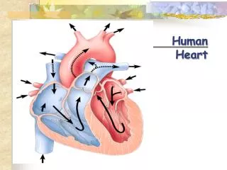
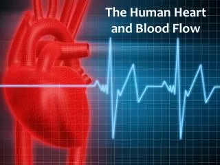
Chambers • 4 Chambers of the Heart. • Two Superior Atria. • Two Inferior Ventricles. • The right hand side of the Heart has a thicker Myocardium because it needs to pump blood at a higher pressure.
Cardiac Cycle • Atrial Systole – Atria contract and the blood flows into the Ventricles. The Semi-Lunar valves are closed. • Ventricular Systole – Ventricles contract and the blood is forced into the Pulmonary Artery and Aorta. The Atrioventricular Valves are forced shut. • Diastole – resting phase, all chambers are relaxed and the blood is filling in the Atria.
Cardiac Muscle Cells • Found on the walls of the heart. • It is resistant to fatigue. • Coronary arteries refresh and supply oxygen to the cells.
Electrical Impulse (II) • The Cardiac muscles need to be able to depolarise without using the nervous system as a source. • The depolarisation must be very quick!
Calcium ions for Contraction • During a contraction, many cross-bridges are continuously made and broken causing them to slide past thick filaments and so shorten the sarcomere. • Tropomyosin (a rod shaped protein) subunits cover myosin head groups on actin fibres.