Download
1 / 66
660 likes | 823 Views
Clinical Pathological Conference Colorectal Cancer. Dr. L. Oliveira – Gastroenterology Dr. C. Walsh – Radiology Dr. F. Halwani - Pathology. OBJECTIVES. Explain the adenoma – carcinoma sequence and rationale behind colon cancer screening.

E N D
Clinical Pathological ConferenceColorectal Cancer Dr. L. Oliveira – Gastroenterology Dr. C. Walsh – Radiology Dr. F. Halwani - Pathology
OBJECTIVES • Explain the adenoma – carcinoma sequence and rationale behind colon cancer screening. • Describe the epidemiology, etiology and risk factors for colonic adenocarcinomas. • Outline the clinical presentation, diagnosis and staging of colonic adenocarcinomas. • Discuss the prevention, management and prognosis of colonic adenocarcinomas. • Summarize the options and guidelines for colon cancer screening and surveillance. • Describe, in general, features of the major hereditary gastrointestinal polyposis syndromes including Hereditary Non‐Polyposis Colorectal Cancer (HNPCC), Familial Adenomatous Polyposis (FAP) and Peutz‐Jeghers Syndrome.
OBJECTIVES • Identify the best radiological imaging modality to screen for colonic polyps. • Identify the best radiological imaging modality to screen for colon cancer • Identify the best radiological imaging modality to stage colon cancer. • Classify polyps into benign and pre‐malignant categories and according to pathological features. • Identify the morphological features of the different types of colonic polyps. • Identify the morphological features of colonic carcinoma.
Question # 1 • All of the following are risk factors for colon cancer except: A. Age B. Hyperplastic polyps C. Family history of colon cancer D. Primary sclerosing cholangitis
Question #2 • If an adenomatous polyp is found and removed on colonoscopy, a surveillance colonoscopy should be done… • A. in 10 years • B. in 2 years • C. in 5 years • D. in 1 year
Question #3 • All of following polyps are benign (non-neoplastic) except: • A. Hamartomatous polyps • B. Serrated polyps • C. Hyperplastic polyps • D. Inflammatory polyps
CASE 1 • 53 yo male referred for weight loss and rectal bleeding • PMHx: healthy • MEDS: none • FAM HX: mother had colon cancer age 56 • HPI: • lost 30lbs in the last 4 months • Decreased appetite • Bowel movements associated with dark blood on and off
CASE 1 • P/E: unremarkable • Labs: Hb 110 (115-155g/L) • Ferritin 10 ug/L
CRC : Diagnosis - Colonoscopy • Colonoscopy has advantage that one can biopsy • In 5 % of cases scope can not be passed to cecum • There is a miss rate associated with colonoscopy • 1/1000 perforation rate. • Risks of sedation • Can be pain during procedure • Greater emphasis being paid to screening of populations at risk and average risk patients
CRC : Clinical Features • Often asymptomatic and present for a long time before symptoms appear • Symptoms relate to tumour size and location Occult Bleeding ( right sided colon cancer ) Obstructive symptoms /change in bowel pattern ( left sided colon cancer ) Rectal Bleeding ( left sided colon cancer ) Weight Loss Abdominal Pain Altered Bowel Pattern Perforation/Abscess Tenesmus/ Incomplete evacuation
CRC : Clinical Features • Cancers of proximal colon tend to grow larger before causing symptoms. May present with occult blood loss and iron deficiency anemia ( fatigue, SOBOE, exertional chest pain) • Cancers of the distal colon tend to have less room to grow and may present with obstruction or rectal bleeding. May have altered bowel pattern, pencil thin stool
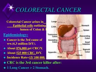
Colon Cancer : Epidemiology • 4th most common cancer ( after prostate, breast, lung) • 10 % of all new cancers • Average individual : 6 % lifetime risk of CRC • 2-3 times increase risk if a first degree relative is affected • Progressive decline in mortality due to earlier detection and better treatment • 70 % of cases are sporadic. ie no family history, no history of IBD.
Colorectal Cancer (CRC) : Epidemiology • 98% Adenocarcinoma. Other ( Lymphoma, Carcinoid, Leiomyosarcoma) • Sporadic : 70% Familial : 25% Inherited : small percentage ( HNPCC, polyposis syndrome) • Risk Factors ?
Colorectal Cancer (CRC) : Risk Factors Disease of developed countries Age : 90% of cases after age 50
Colorectal Cancer (CRC) : Risk Factors Risk Factors • Environmental factors play a minor role high fat low fibre high red meat low calcium low folate • Adenomatous Polyps
Colorectal Cancer (CRC) : Risk Factors • Family History : First degree relative with colorectal cancer • Polyposis Syndromes ex Familial Adenomatous Polyposis ( FAP) Hereditary Non Polyposis Colorectal Cancer (HNPCC) Juvenile Polyposis Syndrome Peutz Jeghers Syndrome • Inflammatory Bowel Disease (IBD) / Primary Sclerosing Cholangitis (PSC) • ASA/NSAID protective
Polyps • Discrete mass of tissue protruding into lumen of colon from normally flat mucosa • Generally asymptomatic • Important characteristic is histology Neoplastic Non Neoplastic • Removal of adenomatous polyps prevents CRC • Closer surveillance of patients who have had adenomas removed
Polyps Can be characterized by gross appearance Sessile vs pedunculated (on a stalk) Removal of adenomatous polyps prevents CRC
Polyps : Classification Non Neoplastic : Benign Hyperplastic – 90% Inflammatory Hamartomatous: Juvenile, Peutz-Jeghers polyps Lymphoid Neoplastic : Characterized by cellular dyplasia Adenomas Tubular, Villous, Tubulovillous, Serrated Malignant polyps
Hyperplastic polyp Serrated architecture
Hamartomatous polyps:1.Juvenile (retention) polyp • Most common childhood polyp • Usually children < 5 years, may occur in adults • 80% in rectum • Commonly presents with rectal bleeding polyps may autoamputate (10%) due to torsion • Usually sporadic; rarely associated with juvenile polyposis syndrome • Not neoplastic by themselves, but may be associated with dysplasia
Hamartomatous polyps:2.Peutz-Jeghers polyp • Hamartomatous polyps that involve the mucosal epithelium, lamina propria, and muscularis mucosa. • May occur singly or multiple in the Peutz-Jeghers syndrome • Syndromic patients have an increased risk of developing GI (2-12%) and extra GI carcinomas ( pancreas, breast, lung, ovary, and uterus) • Solitary polyps do not have malignant potential, occur in colon • Syndromic polyps occur mostly in jejunum intussusceptions
Peutz-Jeghers polyp Arborizing MM (looks like a tree)
Neoplastic polyps • Polypoid areas of epithelial dysplasia • Adenoma = low grade dysplasia • Risk of invasive colorectal adenocarcinoma in the adenoma depends on size: <1% if < 1 cm vs. 10% if > 2 cm; higher risk if villous component • The risk of cancer is high (40%) in sessile villous adenomas > 4 cm • Risk of subsequent carcinoma is related to presence of 3 or more polyps, location at proximal or transverse colon
Tubular adenoma Raised lesion, looks like a berry, protruding in the lumen,attached to a stalk
Tubular adenoma Head of polyp, covered by dysplastic mucosa-with LGD Stalkofpolyp, covered by normal non dysplastic mucosa
VILLOUS ADENOMA Large mass, with a broad, sessile base
VILLOUS ADENOMA Long, slender villi, covered by dysplastic mucosa –with LGD
Malignant Potential of Adenomatous Polyps Nearly all CRC arises from adenomas, but minority ( 5 % or less) of adenomas progress to cancer Prevalence: 25-30 % of people at age 50 will have adenomas • Size Diminutive polyp ( <5 mm) . Low risk of progression • Histologic Type • Degree of Dysplasia Adenoma with Advanced Pathology ( >1 cm, villous, or high grade dysplasia)
Adenoma – Carcinoma Hypothesis • Accumulation of genetic alterations leads to adenoma progression • On average, felt to be 10 years from initiation of adenoma to • progression to adenocarcinoma • APC gene mutation is the critical first step in adenoma formation. • ( Epithelial cells lose function of both APC alleles ). • In Familial Adenomatous Polyposis (FAP) , one APC gene • is inherited in a mutated form
Colon adenocarcinoma Right side carcinoma- can get really large before symptoms, cecum very distensible Left side carcinoma – usually presents with obstruction
The single most important prognostic indicator : extent of the tumor at the time of diagnosis (TNM stage) T- tumor N - lymph node M - metastasis
Pathologic Staging (pTNM)- AJCC 2002 - Primary Tumor (pT) • pTX: Cannot be assessed • pT0: No evidence of primary tumor • pTis: Carcinoma in situ, intraepithelial (no invasion) • pTis: Carcinoma in situ, invasion of lamina propria • pT1: Tumor invades submucosa • pT2: Tumor invades muscularis propria • pT3: Tumor invades through the muscularis propria into the subserosa or the nonperitonealized ericolic or perirectal soft tissues • pT4: Tumor directly invades adjacent structure
Can you identify the layers of colon wall? Can you stage depth of invasion (pT)? 1 1
Regional Lymph Nodes (pN) • pNX: Cannot be assessed • pN0: No regional lymph node metastasis • pN1: Metastasis in 1 to 3 lymph nodes • pN2: Metastasis in 4 or more lymph nodes
Clinical staging of CRC TNM Stage Groupings Astler-Coller Stage Dukes Stage 0 Tis N0 M0 N/A N/A Stage I T1 N0 M0 Stage A A T2 N0 M0 Stage B1 A Stage IIA T3 N0 M0 Stage B2 B Stage IIB T4 N0 M0 Stage B3 B Stage IIIA T1,T2 N1 M0 Stage C1 C Stage IIIB T3,T4 N1 M0 Stage C2,C3 C Stage IIIC Any T N2 M0 Stage C1,C2,C3 C Stage IV Any T Any N M1 Stage D D
CRC : Natural history • Start as epithelial lesions • Become invasive, spreading to lymphatics and vascular channels • Rectal cancers tend to progress to involve local structures • Generally , spread to regional and distant lymph nodes, peritoneum, liver via portal circulation and then lung
CRC : Staging • Physical, Bloodwork ( liver enzymes, CEA), CXR, CT abdo / pelvis. • CEA not useful for screening. High CEA at diagnosis is a marker of poor prognosis. CEA useful for follow up • Localized — confined to the primary site (TNM stage I or II ) • Lymph node involvement (TNM stage III) • Distant metastases (TNM stage IV) • Rectal cancers generally have a worse prognosis than colon cancers 5 year survival Stage I (T1-2N0) — 93 percent Stage IIA (T3N0) — 85 percent Stage IIB (T4N0) — 72 percent Stage IIIA (T1-2 N1)— 83 percent Stage IIIB (T3-4 N1) — 64 percent Stage IIIC (N2) — 44 percent Stage IV — 8 percent
CRC : Treatment Exclude synchronous lesion ( 2 distinct primary tumours. 5 % of cases) Surgery is the treatment of choice where possible Generally, adjuvant chemo for Stage 3 ( and selected patients with stage 2 disease) Chemotherapy for metastatic stage 4 disease Consider adjuvant radiation for rectal cancers to reduce local recurrence
CRC : Treatment Palliative stenting for obstructive tumours
CRC : Screening Removal of Polyps can prevent colon cancer Early detection improves prognosis Fecal Occult Blood Testing /Fecal Immunochemistry (FIT) every 1-2 years FOBT is inexpensive and non invasive FOBT testing will reduce CRC mortality FOBT is not a good test to detect polyps Many false positives occur. Only 2 % of positive tests due to cancer
CRC : Screening Flexible Sigmoidoscopy every 5 years Flexible Sigmoidoscopy and FOBT every 5 years Colonoscopy every 10 years starting at age 50 Radiology CT Colonography DCBE every 5 years