Download
1 / 87
880 likes | 1.26k Views
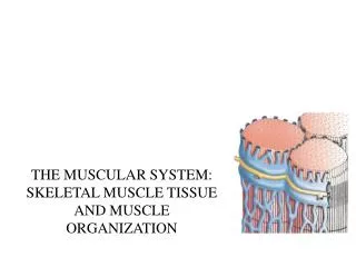
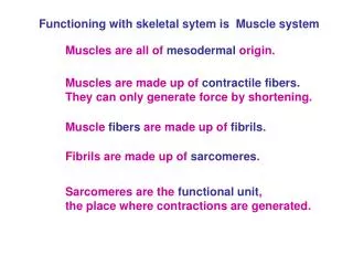
Muscle System B. Ch 9. Sarcolemma. Cell membrane of the muscle fiber Responds to motor neurons Conducts contraction signals. Sarcoplasm. Muscle cytoplasm Contains organelles Mitochondria High concentration Nuclei Multiple Peripheral Transverse tubules Sarcoplasmic reticulum

E N D
Muscle System B Ch 9
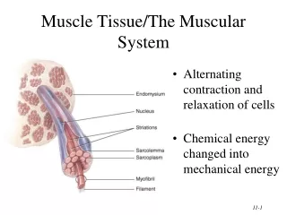
Sarcolemma • Cell membrane of the muscle fiber • Responds to motor neurons • Conducts contraction signals
Sarcoplasm • Muscle cytoplasm • Contains organelles • Mitochondria • High concentration • Nuclei • Multiple • Peripheral • Transverse tubules • Sarcoplasmic reticulum • Myofibrils
Transverse Tubule • Invagination of sarcolemma into the cell • Communicate with all myofibrils • Conducts action potential to cell interior • Distributes contraction signals throughout cell • Communication efficiency • Coordination of muscle contraction • Fluid filled • Specialized interaction with endoplasmic reticulum • Sarcoplasmic reticulum
Sarcoplasmic Reticulum • Specialized Endoplasmic Reticulum • Stores & releases calcium • Terminal Cisternae • Enlarged region of SR • Stores & releases the majority of Ca+ • Connects with T-tubule to form Triad • Arrangement of SR & T-tubule
Myofibril Structure • Protein filaments & motor proteins used in muscle contraction • Myofilament • bundles of protein filaments • Highly organized microfilaments • Work in conjunction with motor proteins • 100-1000+ of myofilaments per myofibril • Change orientation to produce muscle contraction • Active unit • Use ATP
Myofilament • 2 types fibers • Actin- thin filaments • Primarily actin protein • Contain active site • Myosin- thick filaments • Primarily myosin protein • Contain myosin heads • Motor proteins- active site • Tropomyosin • Troponin
Myofilament Organization • Parallel overlapping arrangement • Organization produces striation pattern • Arranged in sarcomeres
Sarcomere • Unit of myofilaments • Comprised of thick & thin fibers • Functional contractile unit • Sarcomere changes length • Protein fibers that make up sarcomere do not change length • 1000s+ make up myofibril • Each contract in coordination with other sarcomeres to create muscle contraction • Distinct organization patterns
Sarcomere Structure • Overlapping fibers with thick filaments in the center & thin filaments on either side • Z line • Protein ends mark boundary of sacromere • Thin filaments attach here • M line • Connect thick filaments • A band • Thick & thin filaments • I band • Thin filaments only • H band • Gap between thin filaments
I band • Isotropic • Thick light outer band • Comprised of only thin filaments • Gets smaller during contraction
A Band • Anisotropic • An= not • Thick dark central band • Comprised of both thick & thin filaments • Remains the same during contraction
Z Line • Aka Z Disc, Zwischen Scheibe • Zwischen= disc Scheibe= inside • Dark, outer boundary of sarcomere • Region of interlocking thin filaments • Move closer together during contraction
H Band • Aka H Zone • Thin, light, middle band • “middle of sarcomere” • Comprised of thick filaments only • Gets smaller during contraction
M Line • Thin, dark, middle line • Region of interlocking thick filaments • Does not change during contraction
Zone of Overlap • Area where thick and thin filaments overlap • Increases during muscle contraction
http://www.youtube.com/watch?v=EdHzKYDxrKc&feature=player_embeddedhttp://www.youtube.com/watch?v=EdHzKYDxrKc&feature=player_embedded
Muscle Contractions • Cumulative shortening of each sarcomere • Actions of myofilaments & motor proteins cause filaments to move against each other in a sliding fashion • Sliding Filament Theory • Coordinated movements • Occur in unique sequence of events triggered by electrical signal
Response Conduction Contraction
Response • Responds to motor neurons • Neuromuscular junction • Site of communication with motor nerves • motor end plates • Motor end plates • High density of receptors • Respond to nerve signal • Neurotransmitters • Ex Ach: Acetylcholine
Conducts • Conducts contraction signals to myofibrils in sarcoplasm • Signal is an electrical current • Action potential • Region of specialization to maximize communication efficiency • Transverse tubule (T Tubule) • Communicates signal to myofibrils
Contraction • Sliding Filament Theory • Contraction a result of coordinated sarcomere shortening • Sarcomere, not filaments change in length • Thin filaments slide inward across thick filaments • Slide over thick filaments • Increase zone of overlap • Produce shortening of sarcomere
Contraction of Sarcomere http://www.youtube.com/watch?v=Ds6f5qeLA8c&feature=related
“Sliding” • Interaction between thin & thick filaments • Active site • Found on thin fibers • Tropomyosin & Troponin • Will interact with myosin heads to create contraction • Connection with myosin forms cross bridge
Myotonia Congenita • Lack of muscle relaxation after contraction • http://www.youtube.com/watch?v=f_3Utmj4RPU&feature=fvw
Muscle Contraction • Uses both chemical & electrical signal • Signal received at sarcolemma and travels to the interior of the cell via T tubules • T Tubules interact with sarcoplasmic reticulum at the cisternae • Trigger release of stored calcium from triad region • Calcium binds with troponin & causes exposure of active site • Myosin cross bridge formed when myosin heads attach to exposed active site • Movement (contraction) • ATP molecule attaches to release cross bridge