Download
1 / 29
541 likes | 1.84k Views
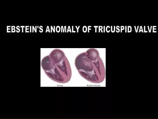
Tricuspid Valve Diseases. M.Sahebjam M.D. Echocardiologist Tehran Heart Center. The forgotten valve. Tricuspid Valve Anatomy. TV annuluss. The tricuspid valve is the most apically (or caudally) placed valve with the largest orifice among the four valves.

E N D
Tricuspid Valve Diseases M.Sahebjam M.D. Echocardiologist Tehran Heart Center
TV annuluss • The tricuspid valve is the most apically (or caudally) placed valve with the largest orifice among the four valves. • The tricuspid annulus is oval-shaped and when dilated becomes more circular. • 20% larger than MV annulus . • Normal TV annulus= 3.0 3.5 cm
Leaflets • the tricuspid valve has three distinct leaflets described as septal, anterior, and posterior. • The septal and the anterior leaflets are larger. • The posterior leaflet is smaller and appears to be of lesser functional significance since it may be imbricated without impairment of valve function.
Leaflets • The septal leaflet is in immediate proximity of the membranous ventricular septum, and its extension provides a basis for spontaneous closure of the perimembranous ventricular septal defect. • The anterior leaflet is attached to the anterolateral margin of the annulus and is often voluminous and sail-like in Ebstein’s anomaly.
Papillary Muscles & Chordae • There are three sets of small papillary muscles, each set being composed of up to three muscles. • The chordae tendinae arising from each set are inserted into two adjacent leaflets. • the anterior set chordae insert into half of the septal and half of the anterior leaflets. • The medial and posterior sets are similarly related to adjacent valve leaflets.
Etiology of Primary Tricuspid Valve Disease • Congenital —Cleft valve generally in association with atrioventricular canal defect —Ebstein’s anomaly —Congenital tricuspid stenosis —Tricuspid atresia • Rheumatic valve disease, generally in association with rheumatic mitral valve disease • Infective endocarditis • Carcinoid heart disease • Toxic (eg, Phen-Fen valvulopathy or methysergidevalvulopathy) • Tumors (eg, myxoma) • Iatrogenic—pacemaker lead trauma • Trauma—blunt or penetrating injuries • Degenerative—tricuspid valve prolapse
Etiology of Secondary or Functional Tricuspid Valve Disease • Right ventricular dilatation • Right ventricular hypertension • Global right ventricular dysfunction resulting from cardiomyopathy, myocarditis, or longstanding right ventricular hypertension with fibrosis • Segmental dysfunction secondary to ischemia or infarction of the right ventricle, endomyocardial fibrosis, arrhythmogenic right ventricular dysplasia
Clinical Presentations • Pure or predominant tricuspid stenosis • Pure or predominant tricuspid regurgitation • Mixed
Tricuspid valve disease—Symptoms • Fatigue • Liver/gut congestion • Right upper quadrant discomfort • Dyspepsia • Indigestion • Fluid retention with leg edema • Ascites
Tricuspid valve disease ausculatory findings • Stenosis : Low-to medium-pitch diastolic rumble with inspiratory accentuation • Regurgitation : Soft, early, or holosystolic murmur Augmented with inspiratory effort (Caravallo’s sign) • Prolapse : Systolic click
Substantial tricuspid regurgitation may exist without the classic ausculatory findings. Thus, clinical evaluation including cardiac auscultation cannot be used to exclude tricuspid valve disease.
Key Diagnostic Features • Mild TR is seen in up to 60% and Moderate TR in up to 15% of healthy individuals. • Mild or worse TR in a valve with thin leaflets, normal coaptation, and normal-appearing supporting structures, suggests regurgitation is physiologic or functional .
Key Diagnostic Features • In carcinoid disease, the leaflets are thickened and retracted with a fixed orifice usually leading to predominant regurgitation and less severe stenosis. • Approximately 30% of patients with MVP have redundancy and prolapse of the tricuspid valve, leading to TR.
PAP based on TR Velocity • Mild increased PAP = 2.6 - 2.9 m/s (27-33 mmhg) • Moderate increased PAP = 3.0 - 3.9 m/s (36-60 mmhg) • Severe increased PAP = 4.0≤ (64 mmhg ≤ )
Tricuspid Valve Stenosis 1/The normal tricuspid inflow velocity is less than 0.5 to 1 m/s, with a mean gradient less than 2 mm Hg. 2/The evaluation of tricuspid valve stenosis with Doppler echocardiography is similar to the method described for mitral stenosis , although the constant of 190 has been proposed of the PHT method . 3/Tricuspid stenosis is considered severe when the mean gradient is 7 mm Hg or more and PHT is 190 milliseconds or longer.
Key Diagnostic Features • TS can be missed on routine TIE because the degree of leaflet thickening may appear subtle, even with significant TS. • Planimetry of the tricuspid valve orifice by two-dimensional images is difficult and unreliable. • A pressure half-time ~ 190 msec suggests severe TS • Valve area is less often used for determining TS severity, but an area < 1.3 to 1.5 cm2 is generally considered significant enough to cause symptoms.