Download
1 / 39
550 likes | 702 Views
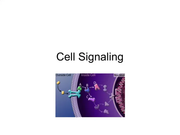
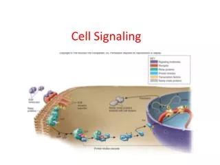
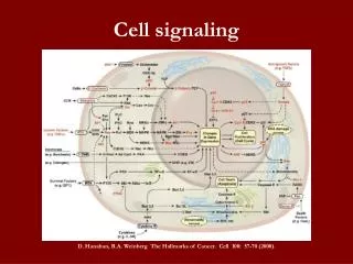
Cell Signaling. I. OVERVIEW. Soluble chemical signals sent from one cell to another are essential for communication The cellular recipient of the signal is known as target cell and binds the signaling molecule via a protein receptor (on cell surface/cytoplasm or nucleus)

E N D
I. OVERVIEW • Soluble chemical signals sent from one cell to another are essential for communication • The cellular recipient of the signal is known as target cell and binds the signaling molecule via a protein receptor (on cell surface/cytoplasm or nucleus) • Binding to receptor initiates signaling cascade of rxns that amplify signal and produce effect.
G-protein Signaling • G proteins are intracellular signaling proteins that are named for their ability to bind to GTP. They also possess GTPase activity. • Two categories of G proteins are described: - heterotrimeric G proteins and - the Ras super-family of G proteins
Rassuperfamily members a.k.a “small G proteins” as they are monomers that resemble one subunit of the heterotrimeric G proteins. • Ras proteins receive their signals from catalytic receptors that have been activated by their ligand. • The overall effects of Ras signaling often involve induction of cell proliferation, cell differentiation, or vesicle transport.
Heterotrimeric G proteins consist of three subunits, α, β, and γ. The signaling process is initiated by ligand binding to receptors linked to G proteins tethered to the inner membrane leaflet. • Activation of the G protein then enables it to regulate a specific membrane-bound enzyme. Products of reactions catalyzed by activated enzymes include second messengers that amplify the signal sent to the cell by the hormone or neurotransmitter that bound to its receptor and acted as the first message.
Many second messengers activate serine/threonine protein kinases, enzymes that phosphorylate their substrates on serine and threonine amino acid residues. Changes in phosphorylation status of target proteins, many of which are enzymes, can alter their activity. The overall result is the biological response of the cell to the hormone or neurotransmitter. The biological response is often the regulation of a biochemical pathway or the expression of a gene.
I. RECEPTORS AND HETEROTRIMERIC G PROTEIN SIGNALING • Many hormones and neurotransmitters have receptors on their target cells that are linked to G proteins. G protein–linked receptors are the most common form of cell surface receptor. • These receptors have extra-cellular hormone-binding regions as well as intracellular portions that interact with the G protein to send the message from the hormone into the cell to evoke a response.
A. G protein–linked receptors • G protein–linked receptors are transmembrane proteins with seven membrane-spanning regions. • Close to 400 distinct G protein–coupled receptors have been identified in humans. • Most are expressed in multiple tissues. Over 90% of them are expressed in the brain. All use the same basic process to stimulate G proteins to regulate the production of second messengers.
B. Signaling mechanism • All heterotrimeric G proteins use the same basic scheme shown for the Gs type of G protein. • An unoccupied G protein–linked receptor does not interact with the G protein in close proximity to its intracellular domain. • Ligand binding to the receptor creates an occupied receptor that undergoes a conformational change and is then able to interact with the G protein. • In response to the receptor binding to the G protein complex, the Gα subunit of the G protein releases GDP and binds GTP. • The G protein is now active and the α subunit dissociates from the β and γ subunits.
The active α subunit then interacts with an enzyme whose function is regulated by the G protein. • Adenylylcyclase is the enzyme activated by Gs protein signaling to have the ability to convert ATP to cyclic AMP (cAMP) and inorganic phosphate (PPi). • cAMP is the second messenger in Gs signaling. The type of G protein that is activated and the second messenger it regulates depend on the ligand, the type of receptor, and the type of target cell. • When hormone is no longer present, the receptor will revert to its resting state. GTP is hydrolyzed to GDP (by the GTPase of the G protein), the enzyme, such as adenylylcyclase, is inactivated, and the α subunit will reassociate with β and γ subunits to stop the signaling process.
III. HETEROTRIMERIC G PROTEINS AND THE SECOND MESSENGERS THEY REGULATE • Distinct members of the heterotrimeric G protein family exist through the association of various forms of the three subunits, α, β, and γ. • At least 15 different α subunits are known. Combinations of different α, β, and γ subunits form the heterotrimeric subunits. • GDP is bound to the α, subunit of the G protein when all three subunits are joined together in the inactive form.
Certain Gα subunits interact with certain enzymes. For example, Gs interacts with adenylylcyclase as described above. • Gα subunits are distinguished from each other by subscripts including s, i, and q (Gαs, Gαi, and Gαq). • The identity of the enzyme determines which second messengers will be produced (or inhibited). Adenylylcyclase and phospholipase C are two enzymes regulated by G proteins that are responsible for regulating messengers with important signaling roles.
A. Adenylyl cyclase • Two different Gα proteins regulate the activity of adenylylcyclase; the Gαs system stimulates its activity while the Gαi inhibits it. • Epinephrine (adrenaline) is a hormone that signals with cAMP as the second messenger. In liver, muscle, and adipose cells, the biological response that results is the breakdown of stored carbohydrates (glycogen) and fat for use as energy. (Glucagon is a hormone that also stimulates glycogen breakdown in liver). • In the heart, the number of beats per minute (heart rate) is increased by this signaling process.
1. Gαs • The active Gα stimulates adenylylcyclase produce the second messenger cAMP. • The enzyme phosphodiesterase converts cAMP to 5′-AMP, ensuring that the amount of cAMP in the cell is low. • cAMP activates cAMP-dependent protein kinase A, known as protein kinase A (PKA). The activation process involves cAMP binding to the regulatory or R subunits of PKA, enabling the release of catalytic or C subunits. Freed C subunits of PKA are active.
PKA phosphorylates its protein substrates, many of which are enzymes, on serine and threonine residues. • Phosphorylation regulates the activity of proteins and enzymes and can lead to intracellular effects. • Protein phosphatases can dephosphorylate the phosphorylated proteins to regulate their activity. • Over time, the Gαs will hydrolyze GTP to GDP to terminate the activation of adenylylcyclase and the production of cAMP.
2. Gαi • When Gαi is activated, it interacts with the active adenylyl cyclase to inhibit its ability to produce cAMP. • In response, PKA will not be activated and its substrates will not be phosphorylated.
B. Phospholipase C • A variety of neurotransmitters, hormones, and growth factors initiate signaling through Gαq (Figure 17.7). • After a hormone binds to its Gq-linked receptor, the intracellular domain of the occupied receptor interacts with Gq. The α subunit of Gq releases GDP and binds GTP. • The α subunit dissociates from the β and γ subunits and then the α subunit activates phospholipase C to cleave the membrane lipid phosphatidylinositol 4,5-bisphosphate (PIP2).
Generation of second messengers in response to Gαq activation of phospholipase C.
The products of this cleavage are inositol 1,4,5-trisphosphate (IP3), which is released into the cytosol, and diacylglycerol (DAG), which remains within the plasma membrane. • IP3 binds to a specific receptor on the endoplasmic reticulum, causing release of sequestered calcium. • Calcium and DAG together activate the calcium-dependent protein kinase named protein kinase C (PKC). • IP3, DAG, and calcium are second messengers in this system.
PKC catalyzes phosphorylation of cellular proteins that mediate cellular responses. Effects of intracellular calcium are mediated by the calcium-binding protein calmodulin (Figure 17.8). • After calcium is released from the endoplasmic reticulum in response to the signaling of hormones or neurotransmitters, the transient increase in intracellular calcium concentration favors formation of the calmodulin-calcium complex. • The calmodulin-calcium complex is an essential component of many calcium-dependent enzymes. Binding of the complex to inactive enzymes results in their conversion to active enzymes.
Calmodulin mediates many effects of intracellular calcium.
IV. RAS G PROTEINS • Ras G proteins are homologous to the α subunits of heterotrimeric G proteins. They do not regulate membrane-bound enzymes or induce the production of second messengers. • Instead, their activation by GTP allows them to initiate a cytoplasmic phosphorylation cascade that termi-nates with activation of gene transcription. • In this signaling scheme, Ras proteins are viewed as relay switches between cell surface receptors and a cascade of serine/threoninekinases that regulate nuclear transcription factors. Such signaling is important in the regulation of cell proliferation.
The aberrant function of Ras proteins may contribute to the malignant growth properties of cancer cells. A. Signaling mechanism • Ras proteins are involved in signaling by certain hormones and growth factors that are ligands of catalytic receptors. • A linear pathway from the cell surface to the nucleus has been described, with Ras acting as an intermediary. • Ligand binding to catalytic receptors can cause phosphorylation of tyrosine residues within the receptors.
The receptor’s phosphotyrosines provide “docking” or binding sites for intracellular adaptor proteins such as SHC and Grb2 that contain regions known as SH2 domains. • Ras-specific guanine exchange factor (GEF) SOS joins the complex, followed by Ras. The SHC-SOS-Ras complex exchanges GTP for GDP on Ras, activating Ras. • Ras-GTP promotes binding and phosphorylation of Raf, a serine protein kinase (also known as MAPKKK for mitogen-activated protein kinasekinasekinase).
A phosphorylation cascade then includes mitogen-activated protein kinases kinases (such as MEK) that phosphorylate and activate mitogen-activated protein kinase (MAPK, aka extracellular signal-regulated kinases or ERK), enabling it to translocate to nucleus where it phosphorylates a transcription factor (such as ELK). • The cascade terminates with transcription of genes for immediate early genes involved in cell division. Hydrolysis of GTP to GDP by RAS terminates the signaling process.
Ras signaling via activation of a cytoplasmic serine/threonine cascade.
This linear pathway is now recognized to be only a part of a very complex signaling circuit in which Ras proteins are involved. • Ras signaling involves a complex array of pathways, where crosstalk, feedback loops, branch points, and multicomponent signaling complexes are seen.
B. Ras mutations and cell proliferation • Mutations in Ras genes result in Ras proteins that cannot hydrolyze GTP to GDP to inactivate the signaling process. • The Ras protein then remains in the active state without stimulation of the receptor and continues to send signals to induce progression through the cell cycle. The result is excessive cell proliferation that can lead to malignancy.
Chapter Summary • G proteins are intracellular signaling proteins named for the ability to bind to and hydrolyze GTP. • Two categories of G proteins are described: heterotrimeric G proteins that regulate second messenger production and Ras superfamily small G proteins. • Heterotrimeric G proteins are composed of α, β, and γ subunits and are activated by ligand binding to G protein–linked receptors.
Active G protein–linked receptors interact with membrane-bound enzymes and regulate their function. • Products of reactions catalyzed by G protein–linked enzymes are second messengers that amplify the signal sent to the cell by the ligand. • Second messengers often regulate the activity of certain serine/threonine protein kinases.
Adenylyl cyclase and phospholipase C are enzymes regulated by G proteins. • Adenylyl cyclase is regulated by Gs proteins that stimulate its activity and Gi proteins that inhibit its activity. • cAMP is the second messenger whose production is regulated by adenylyl cyclase. cAMP activates PKA. • Phospholipase C is activated by Gq proteins that stimulate its activity to cleave the membrane lipid PIP2. • IP3 and DAG are products of this cleavage and are the second messengers.
IP3 induces the release of calcium from the endoplasmic reticulum. • Calcium and DAG activate PKC. • Calcium binds to calmodulin which regulates the activity of other proteins. • The GTP-binding protein Ras is an intermediary in signaling via some catalytic receptors. • Activated Ras can stimulate the MAP kinase cascade of serine/threoninephosphorylations that can result in stimulation of gene transcription. • Ras signaling is involved in the stimulation of cell proliferation. • Mutations in Ras can cause unregulated cell division and malignancy.