Download
1 / 31
310 likes | 318 Views
This chapter explores the structure and function of DNA, including the sugar-phosphate backbone, nitrogenous bases, and the process of DNA replication. Evidence supporting DNA as the genetic material is discussed, along with the experiments conducted by Hershey and Chase. The discovery of the double-helix structure by Watson and Crick is also explained.

E N D
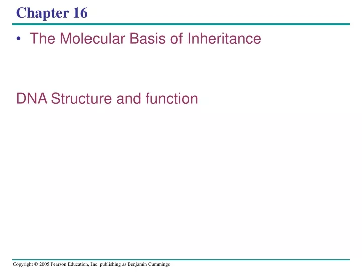
Chapter 16 • The Molecular Basis of Inheritance DNA Structure and function
Sugar-phosphate backbone Nitrogenous bases 5 end CH3 O– 5 O H CH2 O P O O 1 4 N O– N H H H H H O 2 3 H Thymine (T) O H H CH2 O O P N O N H O– H N H H H N N H H Adenine (A) H H O H N CH2 O O P H O O– N H N H H H O H Cytosine (C) O 5 H CH2 O P N O O O 1 4 O– H N H Phosphate H H N 2 H 3 DNA nucleotide N H OH N H Sugar (deoxyribose) 3 end H Figure 16.5 Guanine (G) Additional Evidence That DNA Is the Genetic Material • Prior to the 1950s, it was already known that DNA • Is a polymer of nucleotides, each consisting of three components: a nitrogenous base, a sugar, and a phosphate group
Franklin’s X-ray diffraction Photograph of DNA (b) (a) Rosalind Franklin Figure 16.6 a, b • Maurice Wilkins and Rosalind Franklin • Were using a technique called X-ray crystallography to study molecular structure • Rosalind Franklin • Produced a picture of the DNA molecule using this technique
EXPERIMENT In their famous 1952 experiment, Alfred Hershey and Martha Chase used radioactive sulfur and phosphorus to trace the fates of the protein and DNA, respectively, of T2 phages that infected bacterial cells. CONCLUSION 3 2 4 1 Agitated in a blender to separate phages outside the bacteria from the bacterial cells. Centrifuged the mixture so that bacteria formed a pellet at the bottom of the test tube. Measured the radioactivity in the pellet and the liquid Mixed radioactively labeled phages with bacteria. The phages infected the bacterial cells. Radioactive protein Empty protein shell Radioactivity (phage protein) in liquid Phage Bacterial cell RESULTS DNA Batch 1: Phages were grown with radioactive sulfur (35S), which was incorporated into phage protein (pink). Phage DNA Centrifuge Radioactive DNA Pellet (bacterial cells and contents) Batch 2: Phages were grown with radioactive phosphorus (32P), which was incorporated into phage DNA (blue). Centrifuge Radioactivity (phage DNA) in pellet Pellet Phage proteins remained outside the bacterial cells during infection, while phage DNA entered the cells. When cultured, bacterial cells with radioactive phage DNA released new phages with some radioactive phosphorus. Figure 16.4 Hershey and Chase concluded that DNA, not protein, functions as the T2 phage’s genetic material. • The Hershey and Chase experiment
Figure 16.1 Life’s Operating Instructions • In 1953, James Watson and Francis Crick shook the world • With an elegant double-helical model for the structure of deoxyribonucleic acid, or DNA
G C A T T A 1 nm C G 3.4 nm C G A T G C T A T A A T T A G C 0.34 nm A T Figure 16.7a, c (a) Key features of DNA structure (c) Space-filling model • Watson and Crick deduced that DNA was a double helix • Through observations of the X-ray crystallographic images of DNA
5 end O OH Hydrogen bond P 3 end –O O OH O A T O CH2 O O P O –O O– O P O H2C O O G C O O CH2 O P O O O –O O– O– O– O P P P O O O H2C O O O O C G O O CH2 O P –O O H2C A T O O CH2 OH 3 end (b) Partial chemical structure 5 end Figure 16.7b
H N O H CH3 N N N N H Sugar N N O Sugar Adenine (A) Thymine (T) H O N H N N N H N Sugar N N N O H Sugar H Figure 16.8 Cytosine (C) Guanine (G)
The Basic Principle: Base Pairing to a Template Strand • Since the two strands of DNA are complementary • Each strand acts as a template for building a new strand in replication
T A A A A T A T T T T A G C G C C C C G G G G C A A T T T T A A A A T T T A A A A T A T T T T A C G G G G C G C C C C G (a) The parent molecule has two complementary strands of DNA. Each base is paired by hydrogen bonding with its specific partner, A with T and G with C. (c) Each parental strand now serves as a template that determines the order of nucleotides along a new, complementary strand. (d) The nucleotides are connected to form the sugar-phosphate backbones of the new strands. Each “daughter” DNA molecule consists of one parental strand and one new strand. (b) The first step in replication is separation of the two DNA strands. Figure 16.9 a–d • In DNA replication • The parent molecule unwinds, and two new daughter strands are built based on base-pairing rules
First replication Second replication Parent cell Conservative model. The two parental strands reassociate after acting as templates for new strands, thus restoring the parental double helix. Semiconservative model. The two strands of the parental molecule separate, and each functions as a template for synthesis of a new, comple- mentary strand. Dispersive model. Each strand of both daughter mol- ecules contains a mixture of old and newly synthesized DNA. Figure 16.10 a–c • DNA replication is semiconservative • Each of the two new daughter molecules will have one old strand, derived from the parent molecule, and one newly made strand (a) (b) (c)
DNA Replication chapter 16 continue • DNA Replication a closer look • DNA: Origins of Replication • Elongating a New DNA strand • Antiparallel Elongation • Priming DNA synthesis • Proteins that assist DNA Replication • DNA Proofreading and Repair • Replicating ends of DNA (Telomeres)
DNA Replication: A Closer Look • The copying of DNA • Is remarkable in its speed and accuracy • More than a dozen enzymes and other proteins • Participate in DNA replication
Getting Started: Origins of Replication • The replication of a DNA molecule • Begins at special sites called origins of replication, where the two strands are separated
Origin of replication Parental (template) strand 0.25 µm Daughter (new) strand 1 Replication begins at specific sites where the two parental strands separate and form replication bubbles. Bubble Replication fork 2 The bubbles expand laterally, as DNA replication proceeds in both directions. 3 Eventually, the replication bubbles fuse, and synthesis of the daughter strands is complete. Two daughter DNA molecules (a) In eukaryotes, DNA replication begins at many sites along the giant DNA molecule of each chromosome. (b) In this micrograph, three replication bubbles are visible along the DNA of a cultured Chinese hamster cell (TEM). Figure 16.12 a, b • A eukaryotic chromosome • May have hundreds or even thousands of replication origins
New strand Template strand 3 end 5 end 3 end 5 end Sugar A T A T Base Phosphate C G C G G G C C A T A T OH P P P P 3 end Pyrophosphate P C C OH 2 P 5 end 5 end Figure 16.13 Elongating a New DNA Strand • Elongation of new DNA at a replication fork • Is catalyzed by enzymes called DNA polymerases, which add nucleotides to the 3 end of a growing strand Nucleoside triphosphate
Antiparallel Elongation • How does the antiparallel structure of the double helix affect replication? DNA Replication animation
DNA polymerases add nucleotides • Only to the free 3end of a growing strand • Along one template strand of DNA, the leading strand • DNA polymerase III can synthesize a complementary strand continuously, moving toward the replication fork
To elongate the other new strand of DNA, the lagging strand • DNA polymerase III must work in the direction away from the replication fork • The lagging strand • Is synthesized as a series of segments called Okazaki fragments, which are then joined together by DNA ligase
4 2 3 1 DNA pol Ill elongates DNA strands only in the 5 3 direction. 3 One new strand, the leading strand, can elongate continuously 5 3 as the replication fork progresses. 5 Parental DNA 5 3 Okazaki fragments The other new strand, the lagging strand must grow in an overall 3 5 direction by addition of short segments, Okazaki fragments, that grow 5 3 (numbered here in the order they were made). 2 3 1 5 DNA pol III Template strand DNA ligase joins Okazaki fragments by forming a bond between their free ends. This results in a continuous strand. Leading strand Lagging strand 3 1 2 Template strand DNA ligase Figure 16.14 Overall direction of replication • Synthesis of leading and lagging strands during DNA replication
Priming DNA Synthesis • DNA polymerases cannot initiate the synthesis of a polynucleotide • They can only add nucleotides to the 3 end • The initial nucleotide strand • Is an RNA or DNA primer
Only one primer is needed for synthesis of the leading strand • But for synthesis of the lagging strand, each Okazaki fragment must be primed separately
7 2 3 6 5 1 4 3 5 3 5 Templatestrand DNA pol III adds DNA nucleotides to the primer, forming an Okazaki fragment. Primase joins RNA nucleotides into a primer. RNA primer 3 5 3 1 5 After reaching the next RNA primer (not shown), DNA pol III falls off. Okazakifragment 3 3 5 1 5 After the second fragment is primed. DNA pol III adds DNAnucleotides until it reaches the first primer and falls off. 5 3 3 2 5 1 DNA pol 1 replaces the RNA with DNA, adding to the 3 end of fragment 2. 5 3 3 5 2 1 DNA ligase forms a bond between the newest DNAand the adjacent DNA of fragment 1. The lagging strand in this region is nowcomplete. 5 3 3 2 5 1 Figure 16.15 Overall direction of replication
Table 16.1 Other Proteins That Assist DNA Replication • Helicase, topoisomerase, single-strand binding protein • Are all proteins that assist DNA replication
Overall direction of replication Lagging strand Leading strand Helicase unwinds the parental double helix. Origin of replication 1 Molecules of single- strand binding protein stabilize the unwound template strands. The leading strand is synthesized continuously in the 5 3 direction by DNA pol III. 2 3 Leading strand Lagging strand OVERVIEW DNA pol III Leading strand 5 Replication fork DNA ligase DNA pol I 3 Primase 2 Parental DNA Lagging strand DNA pol III 1 Primer 3 Primase begins synthesis of RNA primer for fifth Okazaki fragment. 4 3 5 4 DNA pol I removes the primer from the 5 end of the second fragment, replacing it with DNA nucleotides that it adds one by one to the 3 end of the third fragment. The replacement of the last RNA nucleotide with DNA leaves the sugar- phosphate backbone with a free 3 end. DNA pol III is completing synthesis of the fourth fragment, when it reaches the RNA primer on the third fragment, it will dissociate, move to the replication fork, and add DNA nucleotides to the 3 endof the fifth fragment primer. DNA ligase bonds the 3 end of the second fragment to the 5 end of the first fragment. 5 6 7 Figure 16.16 • A summary of DNA replication
The DNA Replication Machine as a Stationary Complex • The various proteins that participate in DNA replication • Form a single large complex, a DNA replication “machine” • The DNA replication machine • Is probably stationary during the replication process
Proofreading and Repairing DNA • DNA polymerases proofread newly made DNA • Replacing any incorrect nucleotides • In mismatch repair of DNA • Repair enzymes correct errors in base pairing
1 2 4 A thymine dimer distorts the DNA molecule. • In nucleotide excision repair • Enzymes cut out and replace damaged stretches of DNA A nuclease enzyme cuts the damaged DNA strand at two points and the damaged section is removed. Nuclease DNA polymerase Repair synthesis by a DNA polymerase fills in the missing nucleotides. 3 DNA ligase DNA ligase seals the Free end of the new DNA To the old DNA, making the strand complete. Figure 16.17
5 End of parental DNA strands Leading strand Lagging strand 3 Last fragment Previous fragment RNA primer 5 Lagging strand 3 Primer removed but cannot be replaced with DNA because no 3 end available for DNA polymerase Removal of primers and replacement with DNA where a 3 end is available 5 3 Second round of replication 5 3 New leading strand New lagging strand 5 3 Further rounds of replication Shorter and shorter daughter molecules Figure 16.18 Replicating the Ends of DNA Molecules • The ends of eukaryotic chromosomal DNA • Get shorter with each round of replication
1 µm Figure 16.19 • Eukaryotic chromosomal DNA molecules • Have at their ends nucleotide sequences, called telomeres, that postpone the erosion of genes near the ends of DNA molecules
If the chromosomes of germ cells became shorter in every cell cycle • Essential genes would eventually be missing from the gametes they produce • An enzyme called telomerase • Catalyzes the lengthening of telomeres in germ cells