Download
1 / 16
170 likes | 1.13k Views
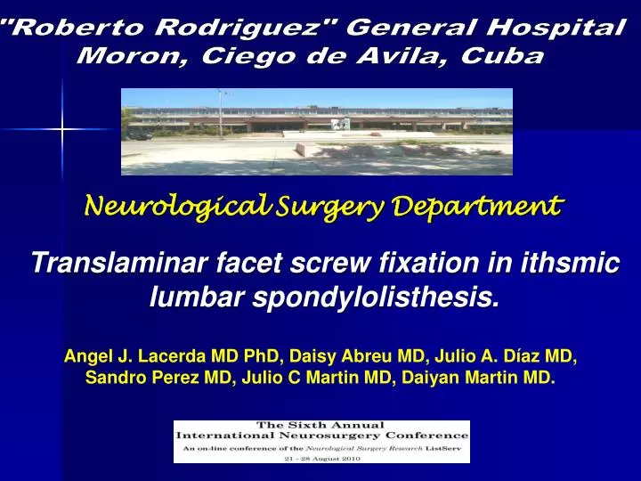
"Roberto Rodriguez" General Hospital Moron, Ciego de Avila, Cuba. Neurological Surgery Department. Translaminar facet screw fixation in ithsmic lumbar spondylolisthesis. Angel J. Lacerda MD PhD, Daisy Abreu MD, Julio A. Díaz MD, Sandro Perez MD, Julio C Martin MD, Daiyan Martin MD.

E N D
"Roberto Rodriguez" General Hospital Moron, Ciego de Avila, Cuba Neurological Surgery Department Translaminar facet screw fixation in ithsmic lumbar spondylolisthesis. Angel J. Lacerda MD PhD, Daisy Abreu MD, Julio A. Díaz MD, Sandro Perez MD, Julio C Martin MD, Daiyan Martin MD.
Introduction: Herbineaux described first spondylolisthesis (1667) in a pregnant woman who had a “tumor” in minor pelvis which was causing obstruction to the delivery. Kilian, Robert, and Lambl described spondylolysis accompanied by spondylolisthesis in the literature in the mid 1800s but development of spondylolisthesis was appreciated only after Naugebauer's anatomic studies in the late 1800s. Wiltse, Macnab, and Newman developed a classification to help outline causes of vertebral translation in an anterior direction.Their categories include the following:
Type I: Congenital spondylolisthesis. Type II: Isthmic spondylolisthesis. Type III: Degenerative spondylolisthesis. Type IV: Traumatic spondylolisthesis. Type V: Pathologic spondylolisthesis.
Isthmic lumbar spondylolisthesis is a defect in the pars interarticularis and occurred when forward translation of lumbosacral vertebra relative to another is of 25%. • Translaminar facet screw fixation is an easy form of lumbar internal fixation but it is less invasive than other techniques. • Indications for this procedure are: • Disabling mechanical lower back pain because of degenerative disk disease or facet joint syndrome. • Symptomatic grade I spondylolisthesis. • The aim of this study was to report the surgical results in our hospital in patients with lumbar ithsmic spondylolisthesis operated on by translaminar facet screw fixation.
Methods: We have conducted a descriptive study about 12 patients operated on neurosurgery department of “Roberto Rodríguez” general hospital, Moron, Ciego de Avila, Cuba, between january 2001 and december 2006, with ithsmic lumbar spondylolisthesis using the translaminar facet screw fixation. Although this technique was described by King in 1948 and Boucher in 1954, the current method of performing the procedure was described by Montesano and colleagues. Spondylolisthesis was evaluated by Meyerding grading system. Results were evaluated by Ebelin scale.
Meyerding grading system classification: Grade 1: 1- 25% slippage Grade 2: 26-50% slippage Grade 3: 51-75% slippage Grade 4: 76-100% slippage Grade 5: Greater than 100% slippage. Grade I lumbar ithsmic spondylolistesis
Surgical procedure: A “meca” position and lumbar midline approach is generally used. Neural microsurgical decompression is performed as indicated. The articular surfaces of the involved facet should not be decorticated to avoid screw purchase weaken. Only the articular cartilaginous end plate is removed.
Only the articular cartilaginous end plate is removed. After that a drill bit is inserted percutaneously and directed toward the base of the spinous process and toward the base of the contralateral transverse process, through a plane across the base of spinous process and lamina and into the facet joint before ending in the transverse process. A small laminotomy allows palpation of the undersurfase of the lamina to avoid drilling into the spinal canal. After that a 4.5 mm standard cortical screw is inserted. The screw length is 50-54mm. The entry point of the second screw is slightly cephalad to avoid crossing the first screw. After screw placement if fusion techcnique will be used the grafts are placed over the graft sites.
Screw inserted Screw inserted
66,67% 33,33% 83,33%
Luque system Translaminar facet screw fixation Transpedicular screw fixation
Conclusions: • There were no complications with this technique. • It is relatively simple and brief technique compared with other stabilization techniques. • Surgical tima is low. • It is biomechanically similar to Luque rectangles. • The facet joint fixating achieves a degree of stabilization that increased the chance for successful fusion in some cases without grafting. • The instrumentation is inexpensive compared with other techniques.