Download
1 / 43
430 likes | 442 Views
Chapter 3 The plasma membrane and membrane potential. Explain how cell membrane constituents function in creating membrane potentials. This will be measured by quiz and exam scores. Review. Membrane structure and composition Cell to cell adhesions Membrane transport New

E N D
Chapter 3The plasma membrane and membrane potential • Explain how cell membrane constituents function in creating membrane potentials. This will be measured by quiz and exam scores.
Review • Membrane structure and composition • Cell to cell adhesions • Membrane transport New • Membrane potentials
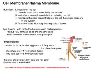
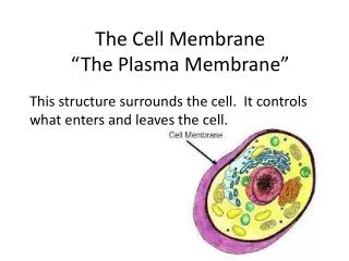
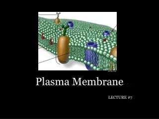
Plasma Membrane • Forms outer boundary of every cell • Controls movement of molecules between the cell and its environment • Joins cells to form tissues and organs • Plays important role in the ability of a cell to respond to changes in the cell’s environment
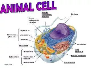
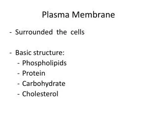
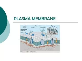
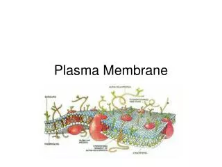
Plasma Membrane Structure • Fluid lipid bilayer embedded with proteins • Most abundant lipids are phospholipids • Also has small amount of carbohydrates • On outer surface only • Cholesterol • Tucked between phospholipid molecules • Contributes to fluidity and stability of cell membrane • Proteins • Attached to or inserted within lipid bilayer
Plasma Membrane Structure ECF Dark line Integral proteins Carbohydrate chain Appearance using an electron microscope Light space Phospholipid molecule Dark line Glycolipid Glycolipid Receptor protein Lipid bilayer Cholesterol molecule Leak channel protein Cell adhesion molecule (linking microtubule to membrane) Carrier protein Microfilament of cytoskeleton Gated channel protein Proteins ICF • Channels • Carrier molecules • Docking marker acceptors • Membrane bound enzymes • Receptor sites • Cell adhesion molecules (CAMs) • Integrin, cadherin • Cell surface markers Fig. 3-3, p. 59
Cell-To-Cell Adhesions • Extracellular matrix • Serves as biological “glue” • Major types of protein fibers interwoven in matrix • Collagen, elastin, fibronectin • CAMs in cells’ plasma membranes • Specialized cell junctions • Desmosomes • Tight junctions (impermeable junctions) • Gap junctions (communicating junctions
Tight junctions Firmly bond adjacent cells together Seal off the passageway between the two cells Found primarily in sheets of epithelial tissue Prevent undesirable leaks within epithelial sheets C Specialized Cell Junctions Desmosomes • Act like “spot rivets” that anchor two closely adjacent nontouching cells • Most abundant in tissues that are subject to considerable stretching Gap junctions • Small connecting tunnels formed by connexons • Especially abundant in cardiac and smooth muscle • In nonmuscle tissues permit unrestricted passage of small nutrient molecules between cells • Also serve as method for direct transfer of small signaling molecules from one cell to the next
Lumen (contains undigested food and potent digestive enzymes) Luminal membrane NO PASSAGE BETWEEN CELLS SELECTIVE PASSAGE THROUGH CELLS Lateral membrane Tight junction Cell 2 Cell 1 Basolateral membrane Blood vessel Epithelial cell lining intestine Fig. 3-5a, p. 63
Membrane Transport • Unassisted membrane transport • Diffusion • Osmosis • Assisted membrane transport • Carrier-mediated transport • Facilitated transport • Active transport
If a substance can permeate the membrane If the membrane is impermeable to a substance Membrane (a) Diffusion occurs (b) No diffusion occurs KEY = Penetrating solute = Nonpenetrating solute Fig. 3-8, p. 66
Area A Area B Area A Area B Diffusion from area A to area B Diffusion from area A to area B Diffusion from area B to area A Diffusion from area B to area A Net diffusion No net diffusion (a) Diffusion (b) Equilibrium KEY = Solute molecule Net diffusion = Diffusion from area A to area B minus diffusion from area B to area A Differences in arrow length, thickness, and direction represent the relative magnitude of molecular movement in a given direction. Fig. 3-7, p. 65
Membrane Transport Factors affecting rate of diffusion collectively make up Fick’s law of diffusion: • Magnitude (or steepness) of the concentration gradient • Permeability of the membrane to the substance • Charge? • Surface area of the membrane across which diffusion is taking place • Molecular weight of the substance • Distance through which diffusion takes place
Membrane Transport • Osmosis • Net diffusion of water down its own concentration gradient
90% water concentration 10% solute concentration 100% water concentration 0% solute concentration (a) Pure water (b) Solution KEY = Water molecule = Solute molecule Fig. 3-9, p. 67
Normal cell volume Intracellular fluid 300 mOsm/L nonpenetrating solutes H2O H2O 300 mOsm/L nonpenetrating solutes 200 mOsm/L nonpenetrating solutes 400 mOsm/L nonpenetrating solutes No net movement of water; no change in cell volume. Water diffuses into cells; cells swell. Water diffuses out of cells; cells shrink. (b) Hypotonic conditions (c) Hypertonic conditions (a) Isotonic conditions Fig. 3-13, p. 71
Membrane Transport Unassisted membrane transport Assisted membrane transport • Carrier-mediated transport • Accomplished by membrane carrier flipping its shape • Can be active or passive • Characteristics that determine the kind and amount of material that can be transferred across the membrane • Specificity • Saturation • Competition
Membrane Transport Types of assisted membrane transport • Facilitated diffusion • Active transport • Vesicular transport
Facilitated diffusion • Substances move from a higher concentration to a lower concentration • Requires carrier molecule • Means by which glucose is transported into cells Carrier protein takes conformation in which solute binding site is exposed to region of higher concentration. 1 Concentration gradient ECF Solute molecule to be transported (High) Carrier protein Plasma membrane Binding site (Low) ICF Direction of transport Transported solute is released and carrier protein returns to conformation in step 1. 4 Solute molecule binds to carrier protein. 2 Carrier protein changes conformation so that binding site is exposed to region of lower concentration. 3 Fig. 3-14, p. 72
Membrane Transport Active transport • Moves a substance against its concentration gradient • Requires a carrier molecule • Primary active transport • Requires direct use of ATP • Secondary active transport • Driven by an ion concentration gradient established by a primary active transport system
Na+ concentration gradient 1 ECF Na+–K+ pump High Na+ Low K+ High-affinity binding site for Na+ Plasma membrane Low-affinity binding site for K+ K+ concentration gradient Low Na+ High K+ ICF 3 Na+ 6 2 Direction of K+ transport 2 K+ 3 Na+ Low-affinity binding site for Na+ High-affinity binding site for K+ Direction of Na+ transport 5 3 2 K+ 4 Active TransportSodium Potassium Pump When open to the ECF, the carrier drops off Na+ on its high-concentration side and picks up K+ from its low-concentration side Stepped Art Fig. 3-16, p. 75
Active Transport • Moves a substance against its concentration gradient. • Primary active transport: • Requires direct use of ATP • Secondary active transport: • Driven by an ion concentration gradient established by a primary active transport • Two types, symportandantiport
Transported solute in low concentration Driving ion in high concentration Driving ion in low concentration Transported solute in high concentration (a) Symport Fig. 3-17a, p. 77
Driving ion in high concentration Transported solute in high concentration Driving ion in low concentration Transported solute in low concentration (b) Antiport Fig. 3-17b, p. 77
Secondary Active transport
Active Transport • Moves a substance against its concentration gradient. • Primary active transport: • Requires direct use of ATP • Secondary active transport: • Driven by an ion concentration gradient established by a primary active transport • Two types, symportandantiport
Carrier-mediated Transport Characteristics • Specificity: Each carrier transports a specific substance or a few closely related compounds. • Saturation: A limited number of carrier binding sites are available. • Transport maximum (Tm): The amount of a substance transported in a given time. • Competition: Several closely related compounds may compete for transport on the same carrier.
Simple diffusion down concentration gradient Carrier-mediated transport down concentration gradient (facilitated diffusion) Rate of transport of molecule into cell Low High Concentration of transported molecules in ECF Fig. 3-15, p. 73
Membrane Transport • Vesicular transport • Material is moved into or out of the cell wrapped in membrane • Active method of membrane transport • Two types of vesicular transport • Endocytosis • Process by which substances move into cell • Pinocytosis – nonselective uptake of ECF • Phagocytosis – selective uptake of multimolecular particle • Exocytosis • Provides mechanism for secreting large polar molecules • Enables cell to add specific components to membrane
What is an excitable cell? Membranes and their potentials are what make cells excitable.
Membrane Potential • Plasma membrane of all living cells has a membrane potential (polarized electrically) • Separation of opposite charges across plasma membrane • Due to differences in concentration and permeability of key ions • Separated charges create the ability to do work like electrons in a battery. • millivolt- 1/1000 volt
Basic Physics • Brownian motion • Electrons protons neutrons • Ohms law E=I*P • Opposites attract, likes repel (hydrophobic/hydrophyllic) • Potential and kinetic energy • Velocity and force, F= MA • Larger mass requires more force to move • Objects in motion stay in motion unless there is friction and drag.
Basic measurements • Volt – unit of charge • mv – 1/1000 volt • Car battery =12V, Cell = -70 mv • Watt = unit of power • Kw = 1000 watts, light bulb = 60 wattsShearon Harris = 900 MW • Ampere • Unit of current • ma = 1/1000 ampere • http://www.osha.gov/SLTC/etools/construction/electrical_incidents/eleccurrent.html
Membrane Potential Which has the greatest membrane potential? B>A B<C
Membrane Potential • Nerve and muscle cells • Excitable cells • Have ability to produce rapid, transient changes in their membrane potential when excited • Resting membrane potential • Constant membrane potential present in cells of nonexcitable tissues and those of excitable tissues when they are at rest • Na+, K+, A-
Membrane Potential • Effect of sodium-potassium pump on membrane potential • Makes only a small direct contribution to membrane potential through its unequal transport of positive ions • The movement of ions and the large negatively charged proteins (A-) generate the potential difference.
Na+ concentration gradient 1 ECF Na+–K+ pump High Na+ Low K+ High-affinity binding site for Na+ Low-affinity binding site for K+ K+ concentration gradient Low Na+ High K+ ICF 3 Na+ 6 2 Direction of K+ transport 2 K+ 3 Na+ Low-affinity binding site for Na+ High-affinity binding site for K+ Direction of Na+ transport 5 3 2 K+ 4 Fig. 3-16, p. 75
ECF Na+–K+ pump (Passive) (Active) Na+ channel K+ channel (Active) (Passive) ICF Fig. 3-23, p. 79
60mv -90mv -70mv • Rp + -70mv • Variable from one cell to another • Poison eliminates this potential • Generated by the imbalance of ions in the intracellular and extracellular spaces.Nernst Equation E=(61) log Co/Ci Table 3-3 p82
E=(61) log Co/Ci For Potassium Ek=(61) log 5mM/150mM For sodium ENa=(61) log 150mM/15mM Co concentration in the ECF Ci concentration in the ICF Used to calculate the contribution of ions to the resting potential of -70mv Nernst equation
EK = -90mv ENa = 60mv ECl = -70mv K and Na drive Cl gradient Resting potential
Neurons and muscle fibers can alter membrane potential to send signals and create motion. Usefulness?