Download
1 / 38
400 likes | 3.5k Views
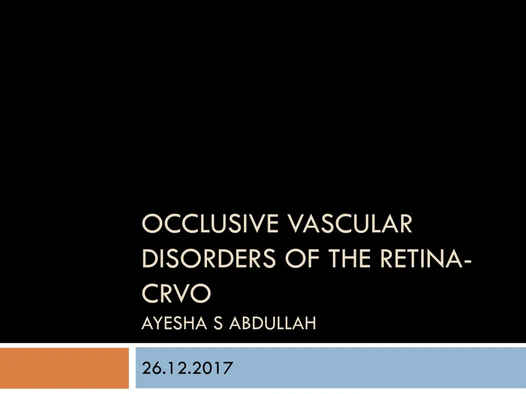
Occlusive vascular disorders of the retina- CRVO Ayesha S abdullah. 26 .12.2017. Review. Learning outcomes. By the end of this lecture the students would be able to Classify occlusive vascular disorders (OVD) of the retina and identify risk factor for these disorders.

E N D
Occlusive vascular disorders of the retina- CRVOAyesha S abdullah 26.12.2017
Learning outcomes By the end of this lecture the students would be able to • Classify occlusive vascular disorders (OVD) of the retina and identify risk factor for these disorders. • Correlate the clinical features of OVD of the retinal with the underlying pathophysiological changes. • Identify CRVO, BRVO, HRVO in given clinical cases or on fundusphotgraphs. • List complications of CRVO and describe prognosis of a given case on the basis of clinical data. • Outline treatment of BRVO and CRVO.
Retinal vein occlusion • Central retinal vein occlusion (CRVO) • Branch retinal vein occlusion (BRVO) • Hemiretinal vein occlusion (HRVO) Retinal arterial occlusion • Central retinal artery occlusion (CRAO) • Branch retinal artery occlusion (BRAO) • Hemiretinal artery occlusion (HRAO)
BRVO HRVO CRVO
Epidemiology • Retinal vein occlusion is the second most common cause of visual loss due to retinal vascular disease • BRVO is the most common type • It is a significant cause of severe visual loss in people over the age of 40 yrs
Pathophysiology • Can you think of some anatomical factors that could predispose retinal veins to occlusion?
With occlusion of the central retinal vein (CRVO) increased venous & capillary pressure Stagnation of blood in the retinal venous system & increased resistance to venous blood flow ischemic damage to the retina increased production of vascular endothelial growth factor (VEGF) neovascularization of the posterior and anterior segment Capillary leakage Complications of RVO Neovascular Glaucoma , NVD, NVE,RD Macular oedema Ischemic Maculopathy
Risk factors Hyperlipid. Clotting. Dis Oral Contracep Hypervisocsity Blood Ocular IOP RV INFLM RVD Vessel wall Age Smoking DM, HTN, AS Infl. Sarcoid. Bechet
CRVO Clinical presentation • Clinical entities • Non –ischemic CRVO (about 75% of cases) • Ischemic CRVO (worse prognosis) • How would you diagnose retinal ischemia?
CRVO Clinical presentation • From painless visual loss to panful blind eye • Decreased vision, metamorphopsia • Visual loss • sudden or gradual, • over a period of days to weeks. • ranges from mild to severe. • Patients can present with transient obscurations of vision initially, later progressing to constant visual loss. • Asymptomatic • Photophobia • Redness of eyes • Painful blind eye
Clinical examination Patients should undergo a complete eye examination, including • visual acuity • pupillary reactions • slit lamp examination of the anterior and posterior segments • undilated examination of the iris….Why? • gonioscopy • Dilated fundus examination
Signs • Visual acuity: (Best-corrected vision acuity) It is one of the important indicators of the final visual prognosis. • Pupillary reactions: • normal/ relative afferent pupillary reflex. If the iris has abnormal blood vessels, the pupil may not react. • Conjunctiva: • Advanced stages may show congestion on conjunctival and ciliary vessels. • Cornea: • Advanced stages may show diffuse corneal edema obscuring the visibility of internal structures.
Signs • Iris • normal/ neovascularization • The anterior chamber angle • it may show neovascularization with open angles and later show total peripheral anterior synechia and closed angles.
Signs • Fundus examination: • Retinal hemorrhages • Dilated tortuous veins • Optic disc edema • Cotton-wool spots • Macular oedema • Late signs: • Neovascularization (NVD, NVE), • optic disc cupping, • optociliary shunt vessels at the disc (a prognostic sign), • pigmentary changes in the macula
Non-Ischemic & Ischemic RVO NICRVO ICRVO • VA Mild to moderate loss • Haemorrhages, CWS, mild macular oedems • Complication- CMO • Severe visual loss • May be painful (NVG) • As of NICRVO with • RAPD • CWS • Complications- NVD, Rubeosis, NVG, CMO, Ischemic maculopathy
Investigations • Risk factors screening (Lab tests) • FFA • Optical Coherence Tomography (OCT) especially for the assessment of macular oedema • Electro-retinogram (ERG)- amplitude of the b-wave is decreased relative to the a-wave
Haemorrhges blocking retinal fluorescence Areas of retinal ischemia
CRVO Prognosis • The prognosis depends upon the reestablishment of patency of the venous system by recanalization, dissolution of clot, or formation of optociliary shunt vessels. • Signs resolve in 6-12 months
TREATMENT • Depends on the type & stage of CRVO • Principles of treatment are • Treat the underlying cause • Monitor • Treatment modalities • Intravitreal corticosteroid and anti-VEGFagents • Dexamethasoneintravitreal implant • Laser photocoagulation
Follow -up • Regular follow –up • “babysit" for these eyes during that period when they are at maximum risk of developing neovascular glaucoma, i.e. first 7-8 months
Prognosis- For nonischemicCRVO • complete recovery with good visual recovery occurs only in about 10% of cases. • 50% of patients will have 6/60 or worse vision. • About 1/3rdof patients convert to ischemic CRVO within 3 years; 15%within the first 4 months. Important to remember
Prognosis- For ischemic CRVO • more than 90% of patients will have 6/60 or worse vision. • About 60% of patients develop ocular neovascularization • About 7-10% of patients can develop CRVO or other type of vein occlusions within either the same eye or the contralateral eye within 2 years. Important to remember
Topics for Test • Diseases of lids • Diseases of cornea • Refractive errors • Uveitits • VR disorder