Download
1 / 54
540 likes | 782 Views
The Skeletomuscular System Bone- brief anatomy, bone formation, disorders Muscular system skeletal- structure, contraction, control smooth muscle cardiac muscle Innervation of muscle. Functions of bone (skeleton) Support and protection Blood cell formation

E N D
The Skeletomuscular System Bone- brief anatomy, bone formation, disorders Muscular system skeletal- structure, contraction, control smooth muscle cardiac muscle Innervation of muscle
Functions of bone (skeleton) Support and protection Blood cell formation Mineral storage (calcium especially) Site for muscle attachmentbody movement
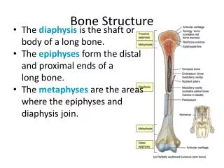
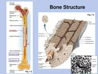
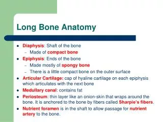
Bones classified by shape: long, short, flat, irregular, round Bone enclosed in periosteum, which is continuous with tendons and ligaments blood vessels in periosteum Epiphysis- ends spongy bone contains red marrow compact bone, articular cartilage Diaphysis- middle compact bone medullary cavity- contains yellow marrow (fat) lined with endosteum (squamous epithelium)
Compact bone osteocytes within lacunae arranged in concentric circles called lamellae This surrounds a central canal; complex is called Haversian system Canaliculi connect osteocytes to central canal and to each other
Prenatal development skeleton is mostly cartilaginous Cartilage cells and then osteoblasts start to deposit minerals Cartilaginous disk (epiphyseal disk) remains in epiphysis Cells eventually stop dividing
Adults continually break down and build up bone Osteoclasts remove damaged cells and release calcium into blood Osteoblasts remove calcium from blood and build new matrix. They become trapped osteocytes
Fracture repair Hematoma- blood clot in space between edges of break Fibrocartilage callus- begins tissue repair Bony callus- osteoblasts produce trabeculae (structural supprt) of spongy bone and replace fibrocartilage Remodeling- osteoblasts build new compact bone, osteoclasts build new medullary cavity
Axial skeleton skull (cranium and facial bones) hyoid bone (anchors tongue and muscles associated with swallowing) vertebral column (vertebrae and disks) thoracic cage (ribs and sternum) Appendicular skeleton pectoral girdle (clavicles and scapulae) upper limbs (arms) pelvic girdle (coxal bones, sacrum, coccyx) lower limbs (legs)
Joints Immovable (synarthoses) bones sutured together by connective tissue: skull Slightly movable (amphiarthoses) connected by fibrocartilage or hyaline cartilage: vertebrae, rib/sternum joint, pubic symphysis Freely movable (diarthroses)- separated ligaments- hold bones together tendons- muscle to bone lined by synovial membrane
Types of freely movable joints Saddle: carpal and metacarpal bones of thumb Ball and socket: shoulder and hip joints Pivot- rotation only: proximal end of radius and ulna Hinge- up and own movement in one plane: knee and elbow Gliding- sliding and twisting: wrist and ankle Condyloid- movement in different planes but not rotations: btw metacarpals and phalanges
Types of movement and examples (with muscles) flexion- move lower leg toward upper extension- straightening the leg abduction- moving leg away from body adduction- movong leg toward the body rotation- around its axis supination- rotation of arm to palm-up position pronation- palm down circumduction- swinging arms in circles inversion- turning foot so sole is inward eversion- sole is out
Elevation and depression- raising body part up or down Aging and bones both bone and cartilage tend to deteriorate cartilage: chondrocytes die, cartilage becomes calcified osteoporosis; bone is broken down faster than it can be built bones get weak and brittle; tend to fracture easily
Skeleton and other systems Skin makes vitamin D which enhances calcium absorption Skeleton stores calcium for muscle contraction, nervous stimulation, blood clot formation Red marrow- site of blood cell formation Calcium levels regulated by parathyroid hormone and calcitonin kidneys (can help provide vitamin D) digestive system (can release calcium into blood)
Growth hormone regulates skeletal growth stimulates cell division in epiphyseal disks in long bones Growth stops when epiphyseal disks are converted to bone When excess growth hormone is produced in childhoodgigantism In adulthood- acromegaly. Bones can’t grow but soft tissue can
When muscle contracts, it shortens and causes movement Skeletal muscles attached to bones by tendons Insertion- attachment to more movable bone Origin- less movable Refer to slide 13. Flexors and extensors act on the same joint to produce opposite actions
Skeletal muscle structure Connective tissue divides muscle tissue into fascicles muscle fibers (myofibers) myofibrils myofilaments (actin and myosin) Each myofiber is formed from several myoblast cells; myofiber cells have multiple nuclei
Muscles appear striated A, I and Z bands appear to change position relative to each other when muscle contracts Each muscle fiber is stimulated by a single axon terminal from a somatic motor neuron Neuromuscular junction- neuron releases acetylcholine at the motor end plate in the sarcolemma
A single somatic motor neuron can produce an axon with several terminal branches. Each stimulates a different muscle fiber. Motor unit- a motor neuron and the muscle fibers it innervates
How muscles contract A bands- thick filaments- myosin I bands- thin filaments- actin “Edges” of A band are darker because thin and thick filaments overlap there H bands- center of A band; lighter because thin filaments do NOT extend there Z bands (disks) define boundaries of the functional unit, or sarcomere
Sliding filament theory of contraction A bands do not decrease in length; I bands do Thin filaments slide past thick filaments How? Cross bridges extending from myosin to actin
Detachment of a cross bridge from actin at end of a power stroke requires a new ATP to bind to myosin ATPase. Rigor mortis at death: no ATP is available ADP remains bound to cross bridges (and thus actin to myosin) Muscles remain stiff until they begin to decompose
Regulation of contraction Actin filament associates with troponin and tropomyosin Tropomyosin blocks the attachment sites in actin for the cross bridges when muscle is realxed To move tropomyosin, troponin interacts with calcium
When Ca 2+ binds troponin, the complex shifts. The cross bridges can now bind to actin. Contraction can continue as long as calcium is bound to troponin.
How is calcium level in cells regulated? Calcium is stored in sarcoplasmic reticulum Released from terminal cisternae by stimulus from motor neuron Transverse tubules are continuous with plasma membrane (sarcolemma). Help conduct action potentials into the muscle fiber.
Relaxation of muscle sarcoplasmic reticulum actively accumulates calcium process involves hydrolysis of ATP (Note: muscle activity requires a lot of ATP)
Movement of skeletal muscles Twitch-rapid contraction and relaxation of fibers Muscle can twitch is response to a single pulse stronger the shock, stronger the twitch graded contraction of whole muscle- depends on number of fibers contracting Summation of twitches- if rapid enough produces tetanus Isometric- muscle exerts tension without shortening Isotonic- shortening does occur
Series-elastic component Tendons have elasticity Elastic recoil helps muscles return to resting length Length-tension muscle is at optimum length for contraction when it is at resting length
Energy requirements of skeletal muscles At rest, most energy obtained from fatty acids Exercise: glycogen and glucose also used ATP is used for: movement of cross bridges pumping of calcium into sarcoplasmic reticulum (i.e., for contraction AND relaxation)
Capacity for aerobic exercise Maximal oxygen uptake (VO2 max) varies by age, size and sex Lactate threshold- percentage of maximal oxygen uptake at which significant increase in lactate is seen (usually 50-70%; higher in athletes) Light exercise: most energy derived from fatty acids At lactate threshold: fatty acids and glucose equally Heavy exercise: over 60% glucose
Oxygen debt- oxygen stores in hemoglobin, myoglobin depleted How is oxygen debt “repaid”? Phosphocreatine helps produce more ATP A lot more phosphocreatine than ATP in muscle cells
Not all muscles have the same contraction speed Slow-twitch- red fibers; lots of myoglobin and blood supply Fast-twitch- fewer capillaries and less myoglobin (white fibers)- anaerobic activity Intermediate fibers- fast twitch but with high oxidative capacity. Resistant to fatigue
A given motor neuron stimulates one type of fiber. Muscles used routinely are mostly smaller with slow-twitch fibers Fatigue accumulation of K+ reduces action potential accumulation of lactic acid lowers pH increased H+ concentration may interfere with other processes?
Effect of endurance training on muscles Increased ability to obtain energy from fatty acids (spare glycogen) Increased myoglobin, mitochondria Lactate threshold is raised Does NOT increase size of muscle (anaerobic training does) e.g., weightlifting- increases thickness of myofibrils and helps build new ones
Alpha and gamma motoneurons can be stimulated simultaneously by upper motor neurons Gamma motoneurons help maintain muscle tone Reflexes- unconscious reaction to muscle stretch (by contraction) monosynaptic- one synapse within CNS Golgi tendon organs- disynaptic reflex monitor tension in tendon Reciprocal innervation- agonist is stimulated, antagonist is inhibited