Download
1 / 38
420 likes | 712 Views
Parasitic Diseases in cultured fishes. By. Dr. Mohamed S. Mohamed Marzouk Professor of Fish Diseases and Management , Faculty of Veterinary Medicine, Cairo University. Parasitic Diseases of cultured fishes. Common Parasites in cultured fishes:. Fish Protozoa (unicellular).

E N D
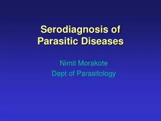
Parasitic Diseases in cultured fishes By Dr. Mohamed S. Mohamed Marzouk Professor of Fish Diseases and Management , Faculty of Veterinary Medicine, Cairo University
Parasitic Diseases of cultured fishes Common Parasites in cultured fishes: • FishProtozoa (unicellular). • Fish Helminthes (multi-cellular). • Fish Parasitic Crustaceans.
Commom protozoa in cultured Fishes: 1. Exteranl cilliated protozoa: • Ichthyophthiriusmultifillis • Chilodonella spp. • Trichodina spp. 2. Internal fish protozoa: • Flagellated Hexamita spp.
Pathogenesis of external protozoa Protozoal invasion Skin cellular irritation 1. Abnormal swimming 2. Excessive mucus Skin cellular destruction 1. Haemorrhages 3. C.T capsules (White spots). 2. Erosions and ulcers
Clinical signs • Abnormal swimming(Flashing, circling, sluggish and itching ). • Surfacing and gasping (Asphyxia). • Excessive skin mucus ( patchy then generalized ). • Pathognomonic white spots (Ichthyophthiriosis). • Skin haemorrhages, erosions and ulcerations. • Emaciation and death.
Diagnosis of External parasitic Diseases 1. History ( new introduced fish, water parameters,……… 2. Clinical signs and lesions. • 3. Demonstration and identification of the causative parasites • Skin and gill mucus scraping (wet mount). • Stained skin and gill mucus smears.
Treatment of External parasitic Fish Diseases • External chemical treatment • Dip treatment • Bath treatment • Flush treatment • Indefinite bath treatment • Non-chemical treatment • Increase water temperature in White Spot disease • Use of ultraviolet radiation in re-circulating system
Types of Chemical treatments 1. Disinfectants: • Pot. Permenganate. • Malachite green. • Formaline. • Na Cl. • Acriflavin. • Methylene blue.
2. Pesticides: • Chlorinated Hydrocarbones. • Organophosphates.
Control of Fish diseases in infected fish farms • In infected earthen ponds: • Drainage • Dryness • Disinfection using quick lime • In infected concrete or fiberglass fish tanks: • Drainage • Disinfection with strong antiseptics
Hexamitiosis • Systemic infection caused by an internal flagellated protozoa Causative protozoa: • Hexamita intestinalis
Mode of infection and transmission: 1. Infection through ingestion. 2. Transmission is from dead fish and contaminated water body. Pathogenesis Hexamita is normal inhabitant of intestine Large numbers Small numbers Systemic form Intestinal destruction No signs Intestinal irritation Hole in head Excess mucus Off food & emaciation
Clinical signs: 1. Off food. 2. Emaciation and tucked up abdomen. 3. Mucus shreds from the vent. 4. High mortality in severe cases. 5. Hole in the head in systemic form (Fistula behind the head exuding white material).
Diagnosis Clinical signs and lesions (Non-confirmative) Laboratory ( Confirmative) Sampling ( Intestinal mucus ) Quantitative (Count/ MF) Qualitative 0 - 5 = Negative 5 – 15 = Mild 15 -30 = Moderate 30 – 100 = Severe More than 100 = Marked
Treatment and control 2. Hexamiticides 1. Expulsion Sulfonamides Saline purgative (MgSO4) • Aresenical (Carbersone). • Murcurial (Calomel). Can be used in food fishes Not to be used in food fishes
Fish Helminthosis Helminthes of fishes Platy helminthes Round worms Trematodes Cestodes Larvae A dults
Fish Trematodes 1. Monogenea 2. Digeanea Adults Gill flukes Skin flukes Encysted metacercaria
Gillflukes 1. Dactylogyridae 2. Cichlidogyridae Cichilidogyrus tilapiae (C. tilapiae) Dactylogyrus vastator (D. vastator)
2 egg sacs Anchor Lernea cyprinicae