Download
1 / 1
10 likes | 253 Views
AMYOPATHIC DERMATOMYOSITIS WITH MASSIVE ECTOPIC CALCIFICATION. A CASE REPORT. E.M. Tinner 1 , I.v. Mühlenen 2 , A. Tyndall 2 , D. Bolz 1 1 Universitätskinderspital beider Basel (UKBB), 2 Felix Platter-Spital Basel. Introduction. Images.

E N D
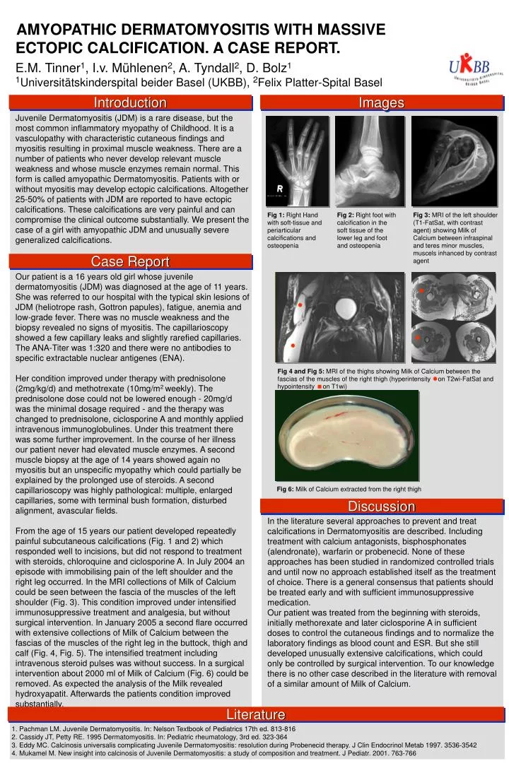
AMYOPATHIC DERMATOMYOSITIS WITH MASSIVE ECTOPIC CALCIFICATION. A CASE REPORT. E.M. Tinner1, I.v. Mühlenen2, A. Tyndall2, D. Bolz1 1Universitätskinderspital beider Basel (UKBB), 2Felix Platter-Spital Basel Introduction Images Juvenile Dermatomyositis (JDM) is a rare disease, but the most common inflammatory myopathy of Childhood. It is a vasculopathy with characteristic cutaneous findings and myositis resulting in proximal muscle weakness. There are a number of patients who never develop relevant muscle weakness and whose muscle enzymes remain normal. This form is called amyopathic Dermatomyositis. Patients with or without myositis may develop ectopic calcifications. Altogether 25-50% of patients with JDM are reported to have ectopic calcifications. These calcifications are very painful and can compromise the clinical outcome substantially. We present the case of a girl with amyopathic JDM and unusually severe generalized calcifications. Fig 1: Right Hand with soft-tissue and periarticular calcifications and osteopenia Fig 2: Right foot with calcification in the soft tissue of the lower leg and foot and osteopenia Fig 3: MRI of the left shoulder (T1-FatSat, with contrast agent) showing Milk of Calcium between infraspinal and teres minor muscles, muscels inhanced by contrast agent Case Report Our patient is a 16 years old girl whose juvenile dermatomyositis (JDM) was diagnosed at the age of 11 years. She was referred to our hospital with the typical skin lesions of JDM (heliotrope rash, Gottron papules), fatigue, anemia and low-grade fever. There was no muscle weakness and the biopsy revealed no signs of myositis. The capillarioscopy showed a few capillary leaks and slightly rarefied capillaries. The ANA-Titer was 1:320 and there were no antibodies to specific extractable nuclear antigenes (ENA). Her condition improved under therapy with prednisolone (2mg/kg/d) and methotrexate (10mg/m2 weekly). The prednisolone dose could not be lowered enough - 20mg/d was the minimal dosage required - and the therapy was changed to prednisolone, ciclosporine A and monthly applied intravenous immunoglobulines. Under this treatment there was some further improvement. In the course of her illness our patient never had elevated muscle enzymes. A second muscle biopsy at the age of 14 years showed again no myositis but an unspecific myopathy which could partially be explained by the prolonged use of steroids. A second capillarioscopy was highly pathological: multiple, enlarged capillaries, some with terminal bush formation, disturbed alignment, avascular fields. From the age of 15 years our patient developed repeatedly painful subcutaneous calcifications (Fig. 1 and 2) which responded well to incisions, but did not respond to treatment with steroids, chloroquine and ciclosporine A. In July 2004 an episode with immobilising pain of the left shoulder and the right leg occurred. In the MRI collections of Milk of Calcium could be seen between the fascia of the muscles of the left shoulder (Fig. 3). This condition improved under intensified immunosuppressive treatment and analgesia, but without surgical intervention. In January 2005 a second flare occurred with extensive collections of Milk of Calcium between the fascias of the muscles of the right leg in the buttock, thigh and calf (Fig. 4, Fig. 5). The intensified treatment including intravenous steroid pulses was without success. In a surgical intervention about 2000 ml of Milk of Calcium (Fig. 6) could be removed. As expected the analysis of the Milk revealed hydroxyapatit. Afterwards the patients condition improved substantially. Fig 4 and Fig 5: MRI of the thighs showing Milk of Calcium between the fascias of the muscles of the right thigh (hyperintensity on T2wi-FatSat and hypointensity on T1wi) Fig 6: Milk of Calcium extracted from the right thigh Discussion In the literature several approaches to prevent and treat calcifications in Dermatomyositis are described. Including treatment with calcium antagonists, bisphosphonates (alendronate), warfarin or probenecid. None of these approaches has been studied in randomized controlled trials and until now no approach established itself as the treatment of choice. There is a general consensus that patients should be treated early and with sufficient immunosuppressive medication. Our patient was treated from the beginning with steroids, initially methorexate and later ciclosporine A in sufficient doses to control the cutaneous findings and to normalize the laboratory findings as blood count and ESR. But she still developed unusually extensive calcifications, which could only be controlled by surgical intervention. To our knowledge there is no other case described in the literature with removal of a similar amount of Milk of Calcium. Literature Pachman LM. Juvenile Dermatomyositis. In: Nelson Textbook of Pediatrics 17th ed. 813-816 Cassidy JT, Petty RE. 1995 Dermatomyositis. In: Pediatric rheumatology, 3rd ed. 323-364 Eddy MC. Calcinosis universalis complicating Juvenile Dermatomyositis: resolution during Probenecid therapy. J Clin Endocrinol Metab 1997. 3536-3542 Mukamel M. New insight into calcinosis of Juvenile Dermatomyositis: a study of composition and treatment. J Pediatr. 2001. 763-766