Download
1 / 46
460 likes | 584 Views
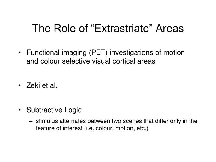
The Role of “Extrastriate” Areas. Functional imaging (PET) investigations of motion and colour selective visual cortical areas Zeki et al. Subtractive Logic stimulus alternates between two scenes that differ only in the feature of interest (i.e. colour , motion, etc.).

E N D
The Role of “Extrastriate” Areas • Functional imaging (PET) investigations of motion and colour selective visual cortical areas • Zeki et al. • Subtractive Logic • stimulus alternates between two scenes that differ only in the feature of interest (i.e. colour, motion, etc.)
The Role of “Extrastriate” Areas • Identifying colour sensitive regions Subtract Voxel intensities during these scans… …from voxel intensities during these scans …etc. Time ->
The Role of “Extrastriate” Areas • result • voxels are identified that are preferentially selective for colour • these tend to cluster in anterior/inferior occipital lobe
The Role of “Extrastriate” Areas • similar logic was used to find motion-selective areas Subtract Voxel intensities during these scans… …from voxel intensities during these scans …etc. STATIONARY STATIONARY MOVING MOVING Time ->
The Role of “Extrastriate” Areas • result • voxels are identified that are preferentially selective for motion • these tend to cluster in superior/dorsal occipital lobe near TemporoParietal Junction • Akin to Human V5
The Role of “Extrastriate” Areas • Thus PET studies doubly-dissociate colour and motion sensitive regions
The Role of “Extrastriate” Areas • V4 and V5 are doubly-dissociated in lesion literature: • achromatopsia (color blindness): • there are many forms of color blindness • cortical achromatopsia arises from lesions in the area of V4 • singly dissociable from motion perception deficit - patients with V4 lesions have other visual problems, but motion perception is substantially spared
The Role of “Extrastriate” Areas • V4 and V5 are doubly-dissociated in lesion literature: • akinetopsia (motion blindness): • bilateral lesions to area V5 (extremely rare) • severe impairment in judging direction and velocity of motion - especially with fast-moving stimuli • visual world appeared to progress in still frames • similar effects occur when M-cell layers in LGN are lesioned in monkeys
The Role of “Extrastriate” Areas • Consider two plausible models: • System is hierarchical: • each area performs some elaboration on the input it is given and then passes on that elaboration as input to the next “higher” area • System is analytic and parallel: • different areas elaborate on different features of the input
How does the visual system represent visual information? How does the visual system represent features of scenes? • Vision is analytical - the system breaks down the scene into distinct kinds of features and represents them in functionally segregated pathways • but… • the spike timing matters too!
Visual Neuron Responses • Unit recordings in LGN reveal a centre/surround receptive field • many arrangements exist, but the “classical” RF has an excitatory centre and an inhibitory surround • these receptive fields tend to be circular - they are not orientation specific How could the outputs of such cells be transformed into a cell with orientation specificity?
Visual Neuron Responses • LGN cells converge on “simple” cells in V1 imparting orientation (and location) specificity
Visual Neuron Responses • LGN cells converge on simple cells in V1 imparting orientation specificity • Thus we begin to see how a simple representation - the orientation of a line in the visual scene - can be maintained in the visual system • increase in spike rate of specific neurons indicates presence of a line with a specific orientation at a specific location on the retina • Why should this matter?
Visual Neuron Responses • Edges are important because they are the boundaries between objects and the background or objects and other objects
Visual Neuron Responses • This conceptualization of the visual system was “static” - it did not take into account the possibility that visual cells might change their response selectivity over time • Logic went like this: if the cell is firing, its preferred line/edge must be present and… • if the preferred line/edge is present, the cell must be firing • We will encounter examples in which these don’t apply! • Representing boundaries must be more complicated than simple edge detection!
Visual Neuron Responses • Boundaries between objects can be defined by color rather than brightness
Visual Neuron Responses • Boundaries between objects can be defined by texture
Visual Neuron Responses • Boundaries between objects can be defined by motion and depth cues
The Feed-Forward Sweep • What is the feed-forward sweep?
The Feed-Forward Sweep • The feed-forward sweep is the initial response of each visual area “in turn” as information is passed to it from a “lower” area • Characteristics: • a single spike per synapse • no time for lateral connections • no time for feedback connections
The Feed-Forward Sweep • The feed-forward sweep is the initial response of each visual area “in turn” as information is passed to it from a “lower” area • What does it mean for an area to be “lower” or “higher”
The Feed-Forward Sweep • Hierarchy of visual cortical areas defined anatomically Dorsal “where”/”how” Ventral “what”
The Feed-Forward Sweep • Hierarchy can be defined more functionaly • The feed-forward sweep is the initial response of each visual area “in turn” as information is passed to it from a “lower” area • Consider the latencies of the first responses in various areas
The Feed-Forward Sweep • Thus the “hierarchy” of visual areas differs depending on temporal or anatomical features • aspects of the visual system account for this fact: • multiple feed-forward sweeps progressing at different rates (I.e. magno and parvo pathways) in parallel • M pathway is myelinated • P pathway is not • signals arrive at cortex via routes other than the Geniculo-striate pathway (LGN to V1) • Will be important in understanding blindsight
The Feed-Forward Sweep • The feed-forward sweep gives rise to the “classical” receptive field properties • tuning properties exhibited in very first spikes • Orientation tuning in V1 • Optic flow tuning in MST • think of cortical neurons as “detectors” only during feed-forward sweep
After the Forward Sweep • By 150 ms, virtually every visual brain area has responded to the onset of a visual stimulus • But visual cortex neurons continue to fire for hundreds of milliseconds!
After the Forward Sweep • By 150 ms, virtually every visual brain area has responded to the onset of a visual stimulus • But visual cortex neurons continue to fire for hundreds of milliseconds! • What are they doing?
After the Forward Sweep • By 150 ms, virtually every visual brain area has responded to the onset of a visual stimulus • But visual cortex neurons continue to fire for hundreds of milliseconds! • What are they doing? • with sufficient time (a few tens of ms) neurons begin to reflect aspects of cognition other than “detection”
Extra-RF Influences • One thing they seem to be doing is helping each other figure out what aspects of the entire scene each RF contains • That is, the responses of visual neurons begin to change to reflect global rather than local features of the scene • recurrent signals sent via feedback projections are thought to mediate these later properties
Extra-RF Influences • consider texture-defined boundaries • classical RF tuning properties do not allow neuron to know if RF contains figure or background • At progressively later latencies, the neuron responds differently depending on whether it is encoding boundaries, surfaces, the background, etc.
Extra-RF Influences • How do these data contradict the notion of a “classical” receptive field?
Extra-RF Influences • How do these data contradict the notion of a “classical” receptive field? • Remember that for a classical receptive field (i.e. feature detector): • If the neuron’s preferred stimulus is present in the receptive field, the neuron should fire a stereotypical burst of APs • If the neuron is firing a burst of APs, its preferred stimulus must be present in the receptive field
Extra-RF Influences • How do these data contradict the notion of a “classical” receptive field? • Remember that for a classical receptive field (i.e. feature detector): • If the neuron’s preferred stimulus is present in the receptive field, the neuron should fire a stereotypical burst of APs • If the neuron is firing a burst of APs, its preferred stimulus must be present in the receptive field
Recurrent Signals in Object Perception • Can a neuron represent whether or not its receptive field is on part of an attended object? • What if attention is initially directed to a different part of the object?
Recurrent Signals in Object Perception • Can a neuron represent whether or not its receptive field is on part of an attended object? • What if attention is initially directed to a different part of the object? Yes, but not during the feed-forward sweep
Recurrent Signals in Object Perception • curve tracing • monkey indicates whether a particular segment is on a particular curve • requires attention to scan the curve and “select” all segments that belong together • that is: make a representation of the entire curve • takes time
Recurrent Signals in Object Perception • curve tracing • neuron begins to respond differently at about 200 ms • enhanced firing rate if neuron is on the attended curve
Feedback Signals and the binding problem • What is the binding problem?
Feedback Signals and the binding problem • What is the binding problem? • curve tracing and the binding problem: • if all neurons with RFs over the attended curve spike faster/at a specific frequency/in synchrony, this might be the binding signal
Feedback Signals and the binding problem • So what’s the connection between Attention and Recurrent Signals?
Feedback Signals and Attention • One theory is that attention (attentive processing) entails the establishing of recurrent “loops” • This explains why attentive processing takes time - feed-forward sweep is insufficient
Feedback Signals and Attention • Instruction cues (for example in the Posner Cue-Target paradigm) may cause feedback signal prior to stimulus onset (thus prior to feed-forward sweep) • think of this as pre-setting the system for the upcoming stimulus • What does this accomplish?
Feedback Signals and Attention • What does this accomplish? • Preface to attention: Two ways to think about attention • Attention improves perception, acts as a gateway to memory and consciousness • Attention is a mechanism that routes information through the brain • It is the brain actively reconfiguring itself by changing the way signals propagate through networks • It is a form of very fast, very transient plasticity
Feedback Signals and Attention • Put another way: • It may strike you as remarkable that a single visual stimulus should “activate” so many brain areas so rapidly • In fact it should be puzzling that a visual input doesn’t create a runaway “chain reaction” • The brain is massively interconnected • Why shouldn’t every neuron respond to a visual stimulus
Feedback Signals and Attention • We’ll consider the role of feedback signals in attention in more detail as we discuss the neuroscience of attention