Download
1 / 41
440 likes | 1.72k Views
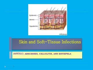
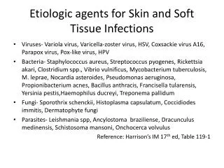
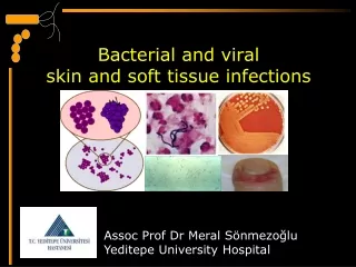
Skin, Soft Tissue, and Bone Infections. Clinical Correlation Series. impetigo. Ecthyma. Erysipelas. Cellulitis. Panniculitis. Necrotizing fasciitis. Considerations in Skin and Soft Tissue Infection. Localization – layer(s) of tissue involved

E N D
Skin, Soft Tissue, and Bone Infections Clinical Correlation Series
impetigo Ecthyma Erysipelas Cellulitis Panniculitis Necrotizing fasciitis
Considerations in Skin and Soft Tissue Infection Localization – layer(s) of tissue involved Localized vs. multifocal; disseminated vs. symmetrical Acute, (bright red, warm, tender) vs. chronic or subacute (dusky red, indurated older eschar or ulcer along with papules) Deep involvement, e.g. muscle (pyomyositis, osteomyelitis, panniculitis Hematogenous vs. exogenous Host factors, exposures
General Rules in Skin Infection • Pustules, tender painful papule or nodule with fluctuance– pyogenic esp. Staph • Spreading erythema, painful , recent onset – Strep, Pasteurella • Bites – cat (Pasteurella), dog (Capnocytophaga), human (Eikenella) • Linear nodules – Tularemia, Mycobacterium, Sporothrix, Nocardia • Vesicles – Herpes, Rickettsialpox • Systemic toxicity, pain out of proportion to appearance – Necrotizing fasciitis • Bullae – Vibrio, Capnocytophaga, Campylobacter • Gangrene – Polymicrobial including Clostridia, enteric GNR • Eschar– Molds, anthrax, tick borne, septicemia • Purpura – Meningococcus, Strep, Staph • Petechiae – Rickettsia, CMV,EBV, HIV (acute)
Classic associations in Skin Infection Finding Organism(s) • Mastectomy Group A strep • Fish Tank M. marinum • Fresh water Aeromonas • Thorn, moss Sporothrix • Neutropenic, moist area Pseudomonas • Neutropenic, tender nodules Candida • Splenectomy Capnocytophaga • Cirrhosis Vibrio • Palms, soles Syphilis, Rickettsia • Eschar Molds, anthrax, Rickettsia • Lymphadenopathy Bartonella, Tularemia
Skin Infection: Geographic Factors • Lyme disease (Erythema chronicum migrans) • Blastomycosis (Ulcerated, verrucous, plaques) • Yersinia pestis (Southwest US) • Coccidioides (Erythema nodosum) • Ehrlichia (RMSF-like illness) • Vibrio, mycobacteria (Gulf coast) • Leishmania (middle east vets)
Fever and Rash: Life threatening Associations Petechial lesions - meningococcal, rickettsial sepsis, TTP* Mucosal involvement – Stevens-Johnson syndrome Bullae – Toxic epidermal necrolysis, Vibrio Purpura – meningococcus, staph, strep, or pneumococus (purpurafulminans) Ecthymagangrenosum– Gram negative sepsis Digital infarcts – Catastrophic APS**, DIC,*** Capnocytophaga, meningococcus • *thrombotic, thrombocytopenic purpura • **antiphospholipid antibody syndrome • ***disseminated intravascular coagulation
Miscellaneous clues to Etiology of Skin infection • Urticaria – hepatitis B (autoimmune reaction) • Slapped cheek, sock and glove purpura– Parvovirus • Hemorrhagic pustules – Neisseria • Nail puncture foot – Pseudomonas • Amoxicillin – EBV • Chronic severe atopy, severe burns – HSV • Intrathoracic or intraabdominal involvement – Actinomycosis, TB • Underlying osteomyelitis– S. aureus, Bartonella • Lung and /or CNS involvement – Nocardia, endemic mycoses, mycobacteria
Fever and Rash: Important Considerations • History must include risk factor assessment – concurrent diseases, medication, travel, occupational/recreational exposure, animals • Thorough exam including entire skin area, mucosa, lymph nodes • Infectious and non infectious diseases can coexist • Skin biopsy for culture and histology rarely contraindicated • Acute retroviral syndrome self-inflicted lesions often not considered
Indications for biopsy, further testing prior to Rx for febrile rash • Chronic or recurrent nature • Ulceration, induration • Failure to respond to seemingly appropriate Rx • Worsening on Rx • Immunocompromised host, trauma, any factor suggesting non infectious cause • Concurrent disease elsewhere, where skin biopsy much less risky than other tissue
Some useful tests for fever and rash Evaluation Test Suspected etiology,clinical setting CXR Mycoplasma, vasculitis Cryptococcal antigen AIDS, transplant and fever CBC with differential Drug reaction, parasite HIV Fever, rash, nodes RPR Palm/sole rash ANA, ANCA Arthralgia, renal disease Serology for RMSF, EhrlichiaPetechiae, headache SPEP Pyodermagangrenosum LFT Urticaria, headache, petechia Blood culture Petechia, toxicity, immunocompromised
Localization of Acute, HematogenousOsteomyelitis Arterial blood flows to blind loop sinusoids
Classification of Osteomyelitis Pathophysiologic Acute vs. chronic Hematogenous vs. contiguous/traumatic Therapeutically Based Medullary Superficial Cortical Localized Diffuse
Osteomyelitis medullary superficial localized diffuse
Symptoms of Osteomyelitis Pain – esp. hematogenous (pediatric, vertebral) may be exquisite or vague may signal complication, e.g. spread to epidural space indistinguishable from sickle cell pain crisis Fever - uncommon
Signs of Osteomyelitis • Erythema, edema, necrosis, bullae, crepitance • Purulence, sinus tract • Non-healing ulcer: cause or consequence • Visible bone (decubitus ulcer) • Non–union of fracture • Separation of components (joint prosthesis) • Elevated WBC, platelets, sedimentation rate , normocytic anemia (of chronic disease) • Radiologic findings
Pathophysiology of Osteomyelitis • Hematogenous – anatomically abnormal bone, prostheses, metaphyses ,vertebral end plate have either increased blood flow a nidus for infection • Contiguous – loss of soft tissue barrier, direct trauma • MSCRAMM – microbial surface components that recognize adhesive matrix molecules • Bacteria adherent to devitalized bone much more resistant to antibiotics
Etiologies of Osteomyelitis • Acute – S. aureus; Salmonella with sickle disease • Contiguous – skin flora; polymicrobial (fecal flora for decubiti, staph strep, anaerobes for diabetes) • Immunocompromised – mycobacteria, fungi, pseudomonas • Prostheses related – Coagulase positive and negative staph, diphtheroids • Vertebral – S. aureus, tuberculosis, endocarditis pathogens
Sequestrum of chronic osteomyelitis Devitalized bone
Medullarry (Hematogenous) Osyeomyelitis Resorbed bone adjacent to growth plate
Osteblastic response to chronic osteomyelitis Hyperdense calcification (involucrum)
MR imaging for osteomyelitis Loss of bone Marrow edema
Diagnostic Pitfalls in Osteomyelitis • Imaging may lag in acute settings • Imaging may distinguish post surgical or traumatic changes • Cultures may reflect surface contaminants • Biopsy may yield sampling error • Nuclear studies may reflect sterile inflammation due to adjacent soft tissue • Neuropathy, decubiti may mask pain • Generally, MR most sensitive, x-rays lag 2 or more weeks behind, negative nuclear studies helpful
Rx of Osteomyelitis • Hematogenous often cured with antibiotic alone • Chronic types esp if cortical or diffuse, prosthesis related, non-union fracture, diabetes related need debridement • Polymicrobial consideration for trauma, contiguous etiology • Usually 6 weeks IV Rx, followed by weeks to months oral agent
Muscle Infection • Quite rare in absence of trauma, ischemia • S. aureus pyomyositis – HIV related in U.S., no obvious risk in tropics • Psoas abscess – relatively common complication of vertebral osteomyelitis (TB, S.aureus) • Parasites – trichinosis • Viral – influenzae B, but not clinically significant • Clostridia – part of fulminant septic picture in setting of underlying malignancy