Download
1 / 34
370 likes | 555 Views
Wet Lab Radiation-induced Apoptosis. Background Equipment Supplies Procedures Lab Demonstrations. Background. Definition Distinctive features of apoptosis Apoptosis vs. necrosis Different stages (detections) of apoptosis. Apoptosis.

E N D
Wet LabRadiation-induced Apoptosis Background Equipment Supplies Procedures Lab Demonstrations
Background • Definition • Distinctive features of apoptosis • Apoptosis vs. necrosis • Different stages (detections) of apoptosis
Apoptosis • A mode of rapid cell death after irradiation • “Programmed” cell death that is potentially a controllable process • Plays an important part in embryogenesis and in tissue regeneration
Relationship between apoptosis and cell kill by radiation Lymphomas 50%-60% Solid tumors 10%-30% Sarcomas < 2-3% Apoptotic death starts 3-5 hours after irradiation Cell loss factor in tumor growth kinetics
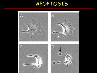
Distinctive features of Apoptosis • Chromatin condensation • Degradation of DNA-ladder form • Cell shrinkage • Scattered throughout a tissue • Inflammation is absent • Activation of a specific program of genes
Necrosis Apoptosis • Accidental death • Severe & sudden injury • ischaemia, physical or • chemical trauma • Cellular and organelle • swelling • Programmed death • Process is more subtle, and • more physiologically • determined. • Cell shrinkage
Necrosis Apoptosis • Random spillage of • cellular content • Inflammatory response • Major site of damage • plasma membrane • Plasma & nuclear membrane blebbing • organelle relocalization & chromatin condensation • Production of membrane enclosed “apoptotic bodies” • Clear by macrophages • No inflammatory response
NECROSIS C B A APOPTOSIS
Detection of Apoptosis by Flow Cytometry • Early stage Annexin V/7-AAD(PI) • Mid stage TUNEL assay • Late stage < Go/G1 DNA content
B C A Apoptosis D F E
Annexin V The translocation of PS precedes other apoptotic processes such as loss of plasma membrane integrity, DNA fragmentation, and chromatin condensation. As such, Annexin V can be conjugated to biotin or to a fluorochrome such as FITC, PE, APC, Cy5, or Cy5.5, and used for the easy, flow cytometric identification of cells in the early stages of apoptosis.
Annexin V Because PS translocation also occurs during necrosis, Annexin V is not an absolute marker of apoptosis. Therefore, it is often used in conjunction with vital dyes such as 7-amino-actinomysin (7-AAD) or propidium iodide (PI), which bind to nucleic acids, but can only penetrate the plasma membrane when membrane integrity is breached, as occurs in the later stages of apoptosis or in necrosis.
Annexin V No Apoptosis = Cell ViabilityCells that are negative for both Annexin V and the vital dye have no indications of apoptosis: PS translocation has not occurred and the plasma membrane is still intact.Early ApoptosisCells that are Annexin V-positive and vital dye-negative, however, are in early apoptosis as PS translocation has occurred, yet the plasma membrane is still intact.
Annexin V Late Apoptosis or Cell DeathCells that are positive for both Annexin V and the vital dye are either in the late stages of apoptosis or are already dead, as PS translocation has occurred and the loss of plasma membrane integrity is observed.When measured over time, Annexin V and a vital dye can be used to monitor the progression of apoptosis: from cell viability, to early-stage apoptosis, and finally to late-stage apoptosis and cell death.
4.72 56.69 93.82 41.84 1.46 2.37 61.22 12.67 37.02 85.25 2.08 1.76
Apoptosis TUNEL ASSAYTdT-mediated dUTP Nick-End Labeling DNA degradation Incorporation of fluorescein-12-dUTP to 3’-OH DNA ends using enzyme Terminal deoxynucleotidyl Transferase (TdT) **** dUTP 5’ 3’ OH
Apoptosis TUNEL Assaywith BrdUrd labelinganti-BrdUrd antibody • Cell number (2 x 10⁶) • Chromatin denaturation • Acid • Pepsin • Separation between neg. and pos. signals • Data analysis • TUNEL assay
30 min 10 min 103 S 102 101 ANTI-BrdUrd ANTIBODY (FITC) G2M 100 G1 10-1 0 10 20 30 40 50 60 0 10 20 30 40 50 60 DNA CONTENT (PI) In vitro: 10 mM BrdUrd In vivo: 10 mg/Kg BrdUrd IP
HL60 cells control 2.0 mg/ml camptothecin Apoptotic cells TUNEL DNA CONTENT
Apoptosis (TUNEL) from Rat Lavage Fluid Control, 11 months Sterling V, 11 months 20.82% 0.97 % Anti-BUDR Antibody (TUNEL) Anti-BUDR Antibody (TUNEL) G1 G1 Cell number Cell number Sub-G1 S G2M S G2M DNA Content DNA Content
Apoptosis Less than Go/G1 DNA content • Gatings • FS : intact cells • DNA (area) vs. DNA (width) • DNA histogram
Equipment • Tissue culture equipment • Sterile tissue culture hood, Incubator (CO2, 37oC, humidified), Microscopes( upright, inverted, dissecting), Pipettes, Hemacytometers (Coulter counter). • Flow cytometer • Radiation source
Supplies • 75% cold (4oC) ethanol • 1x phosphate buffered saline (PBS) • RNase solution in PBS (1 mg/ml) • Propidium iodide solution (20 mg/ml) • APO-BRDU kit, AU1001, Phoenix Flow • Annexin V/PI kit, PF032, Calbiochem • 15 ml conical centrifuge tubes • Pipettes (1ml, 5 ml) • Sample tubes (Falcon 35 2058, 35 2235-filter) • 100 MB Mac formatted zip-disks
Procedures: sample preparation for PI staining(<Go/G1) • Cell permeablization • fresh cells: hypotonic buffers, detergents • fixed cells: 75% EtOH overnight 1x10⁶ cells in 3 ml EtOH • RNase: 1 mg/ml 30 min at room temp. • DNA/PI ratio: 20 mg/ml for 1x10⁶ cells • Cell concentration (1x10⁶/ml)
Procedures: TUNEL assay 10 Gy TdT + BrdU Anti-BrdU antibody
Procedure: Annexin V/PI(7AAD) • Resuspend at least 0.5 x 10⁶ cells in 12x75mm tubes at a concentration of 106 cells/ml. • Add 2ml of cold PBS to tubes. • Centrifuge for 7 minutes at 1,000rpm. • Resuspend pellet in 2ml of cold PBS. • Centrifuge for 7 minutes at 1,000rpm. • Resuspend cells in 0.1ml of 1x binding buffer. • Add 10µl of FITC-conjugated annexin V and 5µl of 7-AAD to tubes. • Gently vortex. • Incubate at room temperature for 15 minutes in the dark. • Add 0.9ml of 1x binding buffer. • Analyze samples within 1 hour of staining.
Lab Demonstrationsroom 3-4151 • Sample measurement • Data analysis • Treatment condition: • 10 Gy, 15 Gy • 0, 6, 24, 48 hr post-irradiation