Download
1 / 38
390 likes | 492 Views
An- Najah National University Faculty of Graduate studies “ Magnatic Resonance Imaging” By Isra Murrar. Outline. Introduction History Alternative Names Uses MRI components How MRI works How to Prepare for the Test How the test will feel Economics of MRI

E N D
An-Najah National University Faculty of Graduate studies “Magnatic Resonance Imaging” By IsraMurrar
Outline Introduction History Alternative Names Uses MRI components How MRI works How to Prepare for the Test How the test will feel Economics of MRI Benefits or advantages Risks or disadvantages
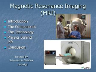
Introduction • Magnetic resonance imaging (MRI) is a test that uses a magnetic field and pulses of radio wave energy to make pictures of organs and structures inside the body. • Because the signal detected by an MRI machine varies depending on the body content and local magnetic properties of a particular area of the body, different tissues or substances can be distinguished from one another in the study image. • In many cases MRI gives different information about structures in the body than can be seen with other imaging method like X-ray, or computed tomography (CT) scan.
History • The development of magnetic resonance imaging (MRI) began with discoveries in nuclear magnetic resonance (NMR) in the early 1900s. At this time, scientists had just started to figure out the structure of the atom and the nature of visible light and ultraviolet radiation emitted by certain substances. • The magnetic properties of an atom's nucleus, which is the basis for NMR, were demonstrated by Wolfgang Pauli in 1924.
History • The first basic NMR device was developed by I. I. Rabi in 1938. This device was able to provide data related to the magnetic properties of certain substances. However, it suffered from two major limitations: • The device could analyze only gaseous materials. • secondly, it could only provide indirect measurements of these materials. • These limitations were overcome in 1945, when two groups of scientists led by Felix Bloch and Edward Purcell independently developed improved NMR devices. These new devices allowed researchers to collect data on many different types of systems. • After further technological improvements, scientists were able to use this technology to investigate biological tissues in the mid 1960s.
History • The use of NMR in medicine soon followed. The earliest experiments showed that NMR could distinguish between normal and cancerous tissue. • Later experiments showed that many different body tissues could be distinguished by NMR scans. • In 1973, an imaging method using NMR data and computer calculations of tomography was developed. It provided the first magnetic resonance image (MRI). This method was consequently used to examine a mouse and, while the testing time required was more than an hour, an image of the internal organs of the mouse resulted. • Human imaging followed a few years later. Various technological improvements have been made since to reduce the scanning time required and improve the resolution of the images. Most notable improvements have been made in the three-dimensional application of MRI.
History • Reflecting the fundamental importance and applicability of MRI in medicine, Paul Lauterbur of the University of Illinois at Urbana-Champaign and Sir Peter Mansfield of the University of Nottinghamwere awarded the 2003 Nobel Prize in Physiology or Medicine for their "discoveries concerning magnetic resonance imaging".
Alternative Names • In the 1970’s the name was changed from NMRI to MRI due to the negative connotations associated with the word “nuclear”. Many patients thought that the exam would expose them to radiation.
Uses Magnetic resonance imaging (MRI) is done for many reasons. It is used to find problems such as • blood vessel diseases • infection • Injuries • tumors
Uses An MRI scan can be done for the: • Head. MRI can look at the brain for tumors, bleeding in the brain, nerve injury, and other problems, such as damage caused by a stroke. MRI can also find problems of the eyes and optic nerves, and the ears and auditory nerves. • Chest. MRI of the chest can look at the heart, the valves, and coronary blood vessels. It can show if the heart or lungs are damaged. MRI of the chest may also be used to look for breast or lung cancer. • Blood vessels. Using MRI to look at blood vessels and the flow of blood through them is called magnetic resonance angiography (MRA) .It can find problems of the arteries and veins, a blocked blood vessel. Sometimes contrast material is used to see the blood vessels more clearly.
Uses • Abdomen and pelvis. MRI can find problems in the liver, gallbladder , pancreas, kidneys. It is used to find tumors, bleeding, infection, and blockage. • Bones and joints. MRI may also be used to tell if a bone is broken when X-ray results are not clear. MRI is done more commonly than other tests to check for some bone and joint problems. • Spine. MRI can check the discs and nerves of the spine for conditions such as spinal tumors.
MRI components • The primary functioning parts of an MRI system include an external magnet, gradient coils, RF equipment, and a computer.
The Primary Magnet • The magnet used to create the constant external magnetic field is the largest piece of any MRI system
There are three types of magnets: • Resistive Magnets • Permanent Magnets • Superconducting Magnets
The resistive magnet • The resistive magnet has many coils of wire that wrap around the bore, through which electrical currents are passed, creating a magnetic field. This particular magnet requires a large amount of electricity to run, but are quite cheap to produce.
The permanent magnet • The permanent magnet is one that delivers a magnetic field, which is always on at full strength and therefore, does not require electricity. The cost to run the machine is low due to the constant magnetic force. However, the major drawback of these magnets is the weight in relation to the magnetic field they produce (more than 7 Tons).
Superconducting Magnets • Most MRI systems use a superconducting magnet, this can be made using various materials, but the basic design involves a coil of conductive wire, a cooling system, and a power supply. The coils are made by wrapping wire, constructed from filaments of a niobium titanium alloy embedded in copper. In order to create a strong enough magnetic field to perform MRI, the coils of wire must have no resistance; therefore they are bathed in liquid helium at a temperature -273.15o C or 0 K! This allows the coils to develop magnetic fields of 1.5 to 3 Tesla (the strength of most medical MRIs), more than 20,000 times stronger than the earth's magnetic field.
The Gradient Coils • In an MRI system, there are typically three sets of gradient coils. Like the primary magnets, each coil is made by winding thin strips of copper or aluminum in a specific pattern. In the MRI system, they are wrapped around the cylinder that surrounds the patient. These magnets are much lower strength compared to the main magnetic field; they may range in strength from 180 gauss to 270 gauss. While the main magnet creates an intense, stable magnetic field around the patient, the gradient magnets create a variable field, which allows different parts of the body to be scanned.
RF equipment • The transmitter and receiver coils are composed of the same type of materials as the gradient coils. They are also constructed much like the main magnet. RF coil is attached to a power source. There are different coils for different parts of the body: knees, shoulders, wrists, heads, necks and so on.
Computer • The computer interprets the data, and creates images that display the different resonance characteristics of different tissue types. We see this as an image of shades of grey-some body tissues show up darker or lighter.
How MRI works • MRI uses properties of hydrogen atoms to distinguish between different tissues within the human body. The human body is composed primarily of hydrogen atoms (63%), other common elements are oxygen (26%), carbon (9%), nitrogen (1%), and relatively small amounts of phosphorus, calcium, and sodium. MRI uses a property of atoms called "spin" to distinguish differences between tissues such as muscle, fat, and tendon.
Placing many protons in a magnetic field, we find that some align anti-parallel and a slight majority aligns parallel. Protons aligned in the parallel orientation are said to be in a low energy state. Protons in the anti-parallel orientation are said to be in a high-energy state.
With a patient in a MRI machine, and the magnet turned on, the nuclei of the hydrogen atoms tend to spin in one of two directions. These hydrogen atom nuclei can transition their spin orientation, or precess, to the opposite orientation. In order to spin the other direction, the coil emits a radiofrequency (RF) that causes this transition (the frequency of energy required to make this transition is specific, and called the Larmour Frequency).
A) The protons spinning in the nature, without an external strong field. The directions of spins are random and cancel out each other. The net magnetization is nearly 0. B) In the presence of a large external magnetic field Bo the spins align themselves either against (high energy state) or along (low energy state). There is a slight abundance of spins aligned in the low energy state.
The signal that is used in creating MRI images is derived from the energy released by molecules transitioning, or precessing, from their high-energy to their low-energy state. This exchange of energy between spin states is called resonance, and thus the name magnetic resonance imaging.
Resonance • The resonance equation shows that the resonance frequency n of a spin is proportional to the magnetic field, Bo, it is experiencing. n = g Bo • Where g is the gyromagnetic ratio. [the ratio of the magnetic moment of a spinning charged particle to its angular momentum].
The RF system has various roles in an MRI machine. First, it is responsible for transmitting the RF radiation that induces the atoms to emit a signal. Next, it receives the emitted signal and amplifies it so it can be manipulated by the computer. RF coils are the primary pieces of hardware in the RF system. They are constructed to create an oscillating magnetic field. This field induces atoms in a defined area to absorb RF radiation and then emit a signal. In addition to sending the RF signal, the coils can also receive the signal from the patient.
The final link in the MRI system is a computer, which controls the signals sent and processes and stores the signals received. Before the received signal can be analyzed by the computer, it is translated through an analog-digital convertor. When the computer receives signals, it performs various reconstruction algorithms, creating a matrix of numbers that are suitable for storage and building a visual display using a Fourier transformer.
How to Prepare for the Test • The Patient may be asked not to eat or drink anything for 4 - 6 hours before the scan. • The Patient should tell doctor if he is afraid of close spaces .He may be given a medicine to help him feel sleepy and less anxious. • Before the test, Patient should tell health care provider if he has: • • Artificial heart valves • • Inner ear implants • • Kidney disease • • Recently placed artificial joints • • Vascular stents • Because the MRI contains strong magnets, metal objects are not allowed into the room with the MRI scanner , for example, items such as jewelry, watches, credit cards, and hearing aids can be damaged.
How the Test Will Feel • An MRI exam causes no pain. If the Patient has difficulty lying still or is very nervous, he may be given a medicine to relax him. • The table may be hard or cold, but Patient can request a blanket. The machine produces loud thumping and humming noises when turned on. Patient can wear ear plugs to help reduce the noise. • An intercom in the room allows Patient to speak to someone at any time. Some MRIs have televisions and special headphones that Patient can use to help the time pass.
Economics of MRI • Standard 1.5 tesla MRI scanners used to cost between US$1 million and US$1.5 million. Standard 3.0 tesla MRI scanners would often cost between US$2 million and US$2.3 million. Construction of MRI suites could cost up to US$500,000 or more, depending on project scope.
Benefits or advantages • MRI systems do not use ionizing radiation, so they can be safely used. • MRI images are clearer and more detailed than with other imaging methods. • MRI enables the detection of abnormalities that might be obscured by bone with other imaging methods. • Another advantage of a MRI is its ability to image in any plane. Other methods of imaging are limited to one plane. • The contrast material used in MRI exams is less likely to produce an allergic reaction
Risks or Disadvantages • MRI scanners are very expensive. • The combination of being put in an enclosed space and the loud noises that are made by the magnets can make some people feel claustrophobic while they are having a MRI scan. • MRI scanners can be affected by movement, making them unsuitable for investigating and less clear. • Although the strong magnetic field is not harmful in itself, medical devices that contain metal may cause problems during an MRI exam.
References • Joseph P. Hornak, “The Basics of MRI”, (1996). • R. Gould, A. Todd and Molly Edmonds, "How MRI Works“, (2010). • John C. Gore, “Magnetic Resonance Imaging”, Elsevier. • Ray Ballinger, “Introduction to MRI”, MRI Tutor physics tutorial, (2013). • B. Disher, L. Lenarduzzi, B. Lewis and J. Teeuwen, “The Physics of MRI”, (2006). • Plewes and Kucharczyk, “Physics of MRI: a primer”, (2012). • Anonick, “The Physics of MRI”, (2009).