Download
1 / 28
890 likes | 4.48k Views
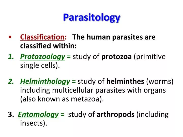
Parasitology. Classification : The human parasites are classified within: Protozoology = study of protozoa (primitive single cells). Helminthology = study of helminthes (worms) including multicellular parasites with organs (also known as metazoa).

E N D
Parasitology • Classification: The human parasites are classified within: • Protozoology =study of protozoa (primitive single cells). • Helminthology = study of helminthes (worms) including multicellular parasites with organs (also known as metazoa). 3. Entomology=study of arthropods (including insects).
Questions form • Type of protozoan (amoeba, ciliate, flagellate: intestinal or blood & tissue, sporozoan) • Disease caused by protozoan • Infective stage • Lab diagnosis (diagnostic stage) • Form of protozoan under the microscope (stage of life cycle) • Vector for the protozoan • Location (site of infection) of the protozoan in the body
1- Entamoeba histolytica • Type of protozoan: Amoeba. • Disease caused by protozoan: Amoebic dysentery. • Infective stage: Cyst. • Diagnostic stage: Cyst or trophozoite in stool. • Forms of the protozoan:Trophozoite or cyst. • Location in the body: Large intestine.
Entamoebahistolytica cyst (picture) Chromatoid bars 4 nuclei
2- Balantidium coli • Type of protozoan: Ciliate. • Disease caused by the protozoan:Balantidial dysentery. • Infective stage: Cyst. • Lab diagnosis (diagnostic stage):Trophozoite or cyst in stool. • Forms of the protozoan:Trophozoite or cyst. • Location in the body: Large intestine.
Balantidium colitrophozoite (H.P) • Largest protozoan. • Trophozoite: oval, covered with cilia, kidney shaped nucleus.
B- Urogenital flagellate Trichomonasvaginalis • Type of protozoan:Urogenital flagellate • Disease caused by protozoan:Trichomoniasis • Infective stage:Trophozoite • Form of protozoan:Trophozoite only. No cyst • Location in the body:Urogenital tract of male (urethra and prostate) and female (vagina) • Lab diagnosis: vaginal swab/discharge or urethral discharge
Trichomonasvaginalistrophozoite (Oil immersion lens) fg: Flagella, nu: nucleus, um: undulating membrane
1- Trypanosomacruzi C- Hemoflagellates • Type of protozoan:Hemoflagellate. • Disease caused by the protozoan:Chagas disease. • Infective stage:Metacyclictrypomastigote. • Form of the protozoan under microscope:Trypomastigote in blood film. • Vector of the protozoan:Bug (Triatoma). • Location of the protozoan in the body:Blood and tissue (heart muscle, liver, spleen, brain). • Diagnostic stage:Trypomastigote in blood film.
Trypanosomacruzitrypomastigote in blood film (Oil immersion lens) Trypomastigote in blood film RBCs
Blood sporozoansPlasmodium spp. • Type of protozoan: Blood sporozoan. • Disease caused by the protozoan: Malaria. • Infective stage:Sporozoites in the saliva of female Anopheles. • Form of the protozoan under microscope: Different stages of the Plasmodium spp. in red blood cells. • Vector of the protozoan: Female anopheles mosquito. • Location of the protozoan in the body: Blood and liver. • Lab diagnosis (diagnostic stage): Different stages of the Plasmodium spp. in red blood cells in blood film.
Different stages of Plasmodium vivax. in blood 1: normal RBCs 2-6: young trophozoite (ring stage) 7-18: trophozoite 19-27: Shizonts 28-29: male and female gametes
Plasmodium spp. Normal RBC’s Different stages of Plasmodium spp. in RBC’s
Tissue sporozoansToxoplasmagondii Toxoplasmagondiitachyzoites (oil immersion lens) are typically crescent shaped with a prominent, centrally placed nucleus.
Toxoplasmagondii Type of protozoan: Tissue sporozoan. Disease caused by the protozoan: Toxoplasmosis. Infective stage:oocyst, cyst, tachyzoites. Form of the protozoan under microscope: Tachyzoites. Location of the protozoan in the body: Different tissues as lung, CNS, heart lymphoid organ and eye. Lab diagnosis (diagnostic stage):Tissue cyst in tonsils and lymph node, X-ray or serologically.
Helminthology Helminths • Helminths (worms)are multicellular parasites. • They are divided into: 1- Flat Worms 2- Round Worms (Platyhelminths) (Nemathelminths) (Flukes) (Tape worms) Class Nematoda Class Trematoda Class Cestoda
Class Trematoda(Flukes) General Characters 1- Flat worms (no body cavity), not segmented, bilaterally flattened. 2-Hermaphrodite. 3- Life cycle show sexual phase (definitive host) and asexual phase (intermediate host). 4- Require one or more intermediate host the 1st intermediate host is a snail.
1- Intestinal FlukeHeterophyesheterophyes • Pear shaped. • Very small size, (2mm). • Oral, ventral, & genital suckers. • Vitelline glands. • 2 testis, 1 ovary. • Simple intestinal ceaca. • (L.P).
Snail: Pirenella conica Eggs: small, operculated, yellowish brown, thin shell (H.P)
Heterophyes heterophyes • Location of adult: Small intestine. • Intermediate host: Primary:Pirenellaconica. Secondary:Bolti and Bouri fish. • Infective stage: Encysted metacercaria. • Mode of transmission: Ingestion of raw or undercooked fish containing encysted metacercaria. • Diagnosis: Eggs in stool. • Disease:Heterophiasis.
2- Liver FlukeFasciola Fasciola hepatica Fasciola gigantica Less prominent shoulders, parallel margins, larger in size (magnifier) 2 prominent shoulders, converging margins, smaller in size (magnifier)
Snail (Lymnaea cailliaudi) Fasciola egg: very large, operculated, yellow, thin shell
Fasciola sp. • Location of adult: Bile duct. • Intermediate host : • Primary : snail Lymnaeatruncatula for F. hepatica and Lymnaeacailliaudi for F. gigantica. • Secondary: leaves of fresh-water plants. • Mode of transmission: Ingestion of raw water-cress containing encysted metacercariae. • Infective stage: Encysted metacercaria. • Diagnosis: Eggs in stool. • Disease:Fascioliasis.