Download
1 / 67
670 likes | 1.81k Views
Arthrology 关节学. 山东大学医学院 解剖学教研室 李振华. Classification . There are two major types of articulations or joint. Continuous joints 直接连结 Fibrous joints 纤维连结 : bones are united by fibrous connective tissue Syndesmosis 韧带连结 Suture 缝 Cartilaginous joints 软骨连结 :

E N D
Arthrology 关节学 山东大学医学院 解剖学教研室 李振华
Classification There are two major types of articulations or joint. • Continuous joints直接连结 • Fibrous joints 纤维连结: bones are united by fibrous connective tissue • Syndesmosis 韧带连结 • Suture 缝 • Cartilaginous joints 软骨连结: bones are united by cartilage • Synchondrosis 透明软骨结合: bones are united by hyaline cartilage • Symphysis 纤维软骨联合: bones are united by fibrocartilage • Synosteosis 骨性结合 • Discontinuous joints间接连结-synovial joints 滑膜关节
Synovial joints滑膜关节 Basic structures • Articular surface关节面: covered by articular cartilage 关节软骨 • Articular capsule 关节囊 • Fibrous membrane纤维膜 • Synovial membrane滑膜 • Articular cavity关节腔: containing a trace of synovial fluid; subatmospheric pressure in it
Accessory structures • Ligaments韧带(lig.): extra- and intracapsular ligaments 囊外(内)韧带 • Articular disc关节盘and articular labrum 关节唇: • Synovial fold滑膜襞and synovial bursa滑膜囊
Terms movements of joints关节运动术语 • Translation 移动 • Flexion and extension 屈和伸 • Adduction and abduction 收和展 • Rotation 旋转 • Medial and lateral rotation 旋内和旋外 • Pronation ans supination旋前和旋后 • Inversion and eversion 足内翻和足外翻 • Circumduction 环转
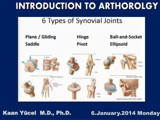
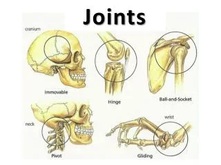
Classification of synovial joints关节的分类 • Uniaxial joints单轴关节: • hinge joints 屈戍关节 • trochoid (pivot) joints车轴关节 • Biaxial joints双轴关节: • ellipsoid joints 椭圆关节 • saddle joints 鞍状关节 • Multiaxial joints多轴关节: • ball-and-socket joint 球窝关节 • plane joints 平面关节
Articulations of Bones of Trunk躯干骨的连结 The vertebral column脊柱consists of 24 vertebrae, the sacrum, and the coccyx.
Joints of the vertebral bodies椎体间的连结 Intervertebral discs 椎间盘 between bodies of adjacent vertebrae, composed of: • Nucleus pulposus髓核, an inner soft, pulpy, highly elastic structure (gelatinous core ) • Annulus fibrosus纤维环 an outer fibrous ring consisting of fibrocartilage
Anterior longitudinal ligament 前纵韧带 • Strong band covering the anterior part of the vertebral bodies and intervertebral discs running from the anterior margin of foramen magnum to the S1~S2 • Maintains stability of the intervertebral disc and prevents hyperextension of the vertebral column Posterior longitudinal ligament后纵韧带 • Attached to the posterior aspect of the intervertebral discs and posterior edges of the vertebral bodies from C2 vertebra to sacrum • Prevents hyperflexion of the vertebral column and posterior protrusion of the discs
Joints of the vertebral arches椎弓间的连结 • Ligamenta flava黄韧带― elastic ligament, unite laminae of adjacent vertebrae, and complete the posterior wall of vertebral canal; tend to prevent hyperflexion of the vertebral column • Interspinal ligament 棘间韧带 • Supraspinal ligament 棘上韧带 • Ligamentum nuchae项韧带 • Intertansverse ligament横突间韧带 • Zygapophysial joint 关节突关节
Atlantooccipital joint寰枕关节 • Between superior articulating surfaces of atlas and occipital condyles • Supported by membrances and ligaments that join occipital bone and atlas • Action ― nodding of head, lateral tilting of head
Atlantoaxial joint寰枢关节 • Three synovial joints between atlas and axis • Laterally, paired joints between articulating facets • Median joint between dens of axis and anterior arch of atlas • Supported by ligaments • apical ligament of dens 齿突尖韧带 • alar ligament 翼状韧带 • transverse ligament of atlas寰椎横韧带 • tectorial membrane 覆膜 • Action ― allow atlas (and head) to pivot on the axis and vertebral column
Normal Curves of vertebral column • Cervical curvature颈曲convex forward • Thoracic curvature胸曲convex backward • Lumbar curvature腰曲 convex forward • Sacral curvature骶曲convex backward Movement of the vertebral column • flexion • extension • lateral flexion • rotation
Thoracic cage 胸廓 Composition Bones ― consists of twelve thoracic vertebrae, twelve pairs of ribs and costal cartilages, and sternum
Joints • Costovertebral joints 肋椎关节 • Joints of costal head 肋头关节 • Costotransverse joints 肋横突关节 • Sternocostal joints 胸肋关节 • Sternocostal synchondrosis of first rib 第一胸肋结合 • Sternocostal joints胸肋关节: • Interchondral joints肋软骨连结: between costal cartilages 8, 9, and 10 to form the costal arch肋弓
General features of thoracic cage • Roughly cone-shape, narrow above and broad below, flattened from before-backwards, longer behind than in front • Inlet of thorax胸廓上口: bounded by upper border of manubrium, first rib, and vertebra T1 • Outlet of thorax胸廓下口: bounded by vertebra T12, 12th and 11th ribs, costal arch and xiphoid process • Infrasternal angle胸骨下角: formed by the costal arch of both side • Intercostal spaces肋间隙: lie between the ribs
Function: • protects the organs in the thoracic cavity and upper abdominal cavity; • plays a vital role in the process of breathing Expiration Inspiration
Joints of skull 颅骨的连结 • Continuous joints: sutures, synchondrosis or synosteosis
Temporomandibular joint 颞下颌关节 • Aticulating surfaces • Mandibular fossa and articular tubercle, above • Head of mandibule, below • Capsule: thin and lax in front and behind; strengthened by the lateral ligament 外侧韧带 • Articular disc: separates surfaces, forming upper and lower compartments within joint • Movement: mandible may be elevated or depressed, protruded or retracted; rotation may also occurs as in chewing( a slight amount of side to side movement is also permitted)
Joints of limbs 山东大学医学院 解剖教研室 李振华
Joints of upper limb Joints of should girdle • Sternoclavicular joint胸锁关节 • Bones: sternal end of clavicle, clavicular notch of sternum, and first costal cartilage • Articular capsule: strong and is reinforced by anterior and posterior sternoclavicular ligaments • An articular disc is attached to the capsule, dividing the joint into two cavities. • Movements: elevation and depression, forward and backward, rotation and circumduction of the acromial end of the clavicle
coracoacromial ligament acromion coranoid process • Acromioclavicular joint 肩锁关节 • Bones: acromion and acromial end of clavicle • Movement: rotation of scapula on clavicle • Coracoacromial arch 喙肩弓 formed by coracoacromial ligament喙肩韧带, coranoid process, and acromion, that prevents the shoulder joint from superior dislocation
Joints of free upper limb ★Shoulder joint肩关节 (ball and socket) • Bones: head of humerus and glenoid cavity of scapula • Capsule: • Thin and lax, especially lower part • Attachments: proximal to glenoid labrum; distal to anatomical neck of humerus, except medially where it is slightly distal to surgical neck • Tendon of long head of biceps brachii passes though the cavity
Accessory structures • Glenoid labrum盂唇: fibrocartilaginous ring on periphery of glenoid cavity • Coracohumeral ligament 喙肱韧带:runs from coracoid process to greater tubercle • Movements: flexion, extension, adduction, abduction, medial and lateral rotation, circumduction
★ Elbow joint肘关节 • Bones: lower end of humerus, upper ends of radius and ulna • Humeroulnar joint 肱尺关节: formed by trochlear of humerus and troclear noch (hinge) • Humeroradial joint肱桡关节: formed by capitulum of humerus and head of radius (ball and socket) • Proximal radioulnar joint桡尺近侧关节: formed by articular circumference of radius and radial notch of ulna • Capsule: thin and lax anteriorly and posteriorly, strongly thickened on either side by collateral ligaments
Ligaments: • Radial collacteral ligament桡侧副韧带: attached to lateral epicondyle and annular ligament of radius • Ulnar collacteral ligament尺侧副韧带: attached to medial epicondyle to medial border of trochlear notch • Annular ligament of radius桡骨环状韧带: attached to anterior and posterior margins of radial notch of ulna, surrounds the head of radius • Movements: flexion and extension, pronation and supination
Joints between radius and ulna • Proximal radioulnar joint桡尺近侧关节 • Distal radioulnar joint桡尺远侧关节: formed by head of ulna, ulnar notch of radius and an articular disc • Interosseous membrane of forearm 前臂骨间膜: a fibrous membrane between the shaft of radius and ulna
Joints of hand ★ Radiocarpal joint桡腕关节(ellipsoid) • Bones • Carpal articular surface of radius and articular disc below the ulna • Proximal row of carpal: scaphoid, lunate, and triquetral bones, but not pisiform • Capsule: lax and strengthened by surrounding ligament • Movements: flexion, extension, adduction, abduction, and circumduction
Intercarpal joints • Carpometacarpal joints: ★Carpometacarpal joint of thumb 拇指腕掌关节 • Bones: trapezium and base of first metacarpal • Movement: flexion, extension, adduction, abduction, and opposition • Intermetacarpal joints • Metacarpophalangeal joints • Interphalangeal joints
Joints of Lower limb Joints of pelvic girdle • Sacroiliac joint 骶髂关节 • Bones: auricular surface of sacrum and ilium • Capsule: very tight and strengthened by ligaments
Vertebropelvic ligaments • Iliolumbal ligament髂腰韧带: runs from transverse process of L5 to the posterosuperior part of iliac crest ★Sacrotuberous ligament 骶结节韧带: runs from lateral margins of sacrum and coccyx to the inner margin of ischial tuberosity ★Sacrospinous ligament 骶棘韧带: runs from ischial spine to lateral margins of sacrum and coccyx • These two ligaments convert the sciatic notches the greater and lesser sciatic foramina 坐骨大、小孔
Pubic symphysis 耻骨联合 • Articulation: symphysial surface and interpubic disc (fibrocartilage) • Ligaments: superior pubic ligament and arcuate pubic ligament • Obturator membrane 闭孔膜 obturator canal 闭膜管
Bony pelvis骨盆 Composition: formed by paired hip bones, sacrum, coccyx, and their articulations • In anatomical position, anterior superior iliac spines and pubic tubercles on same vertical plane, while the tip of coccyx and superior border of pubic symphysis on same horizontal plane • Terminal line界线: formed by promontory of sacrum, arcuate line, pectin of pubis, pubic tubercle, upper border of pubic symphysis • Two portions: a greater pelvis and a lesser pelvis
Lesser pelvis小骨盆 • pelvic inlet骨盆上口(terminal line): • Pelvic outlet 骨盆下口: formed by tip of coccyx, sacrotuberous ligament, ischial tuberosity, ramus of ischium, inferior ramus of pubic, symphysis • Pelvic cavity • Pubic arch, subpubic angle
Main difference between male and femal pelvis Male Female Pelvic inlet Pelvic outet Pelvic cavity Pubic arch 90~1000 70~750
Joints of free lower limb ★ Hip joint 髋关节 • Bones: acetabulum and femoral head • Articular capsule attachments • Above: margins of acetabulum and transverse acetebular ligament • Below: in front to intertrochanteric line; behind, to the neck of femur above 1 cm above the intertrochanteric crest
Acetabulum labrum Ligament of head of femur Transverse acetebular lig. • Accessory structures • Acetabulum labrum髋臼唇; transverse acetebular ligament髋臼横韧带 • Ligaments • Iliofemoral lig. 髂股韧带 • Ligament of head of femur 股骨头韧带 • Pubofemoral lig. 耻股韧带 • Ischiofemoral ligament 坐股韧带 • Zona orbicularis轮匝带 • Movement: flexion, extention, adduction, abduction, medial and lateral rotation, circumduction
Pubofemoral lig. Iliofemoral lig. Ischiofemoral lig. Zona orbicularis
★Knee joint 膝关节 • Bones: lower end of femur, upper end of tibia and patella • Articular capsule: superapatellar bursa髌上囊, deep infrapatellar bursa髌下深囊, ala folds翼状襞