Download
1 / 23
240 likes | 332 Views
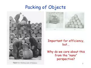
Packing Quality of Atoms in Proteins. Madhuwanti Vaidya Advisor: Herbert Edelsbrunner. Volume of union of spheres. Sum of Volume of spheres. Volume of space. Volume of space. Introduction. Packing Density. p = Covering Density. c =. Gap in Packing and Covering.

E N D
Packing Quality of Atoms in Proteins Madhuwanti Vaidya Advisor: Herbert Edelsbrunner BioGeometry – Duke University
Volume of union of spheres Sum of Volume of spheres Volume of space Volume of space Introduction • Packing Density. p = • Covering Density. c= BioGeometry – Duke University
Gap in Packing and Covering • spheres do not overlap p 1. • spheres cover entire space c 1. Atoms in proteins overlap but do not cover the entire space. Technically neither definitions apply to proteins, though the term ‘packing density’ is commonly used. BioGeometry – Duke University
Why measure packing density in proteins ? • Interior of a protein is tighter packed as compared to exterior. • Protein-protein interaction surface, in a protein-protein complex, consists of regions of high and low packing density. • Hot spots are more likely to be located in regions with higher packing density. BioGeometry – Duke University
Volume of atom Volume of Voronoi cell Current methods • Voronoi volume method. (Richards F. M. 1974) Packing Density = • Occluded surface method. (Fleming et. al. 1995) Packing Density = 1 - PPp BioGeometry – Duke University
Current methods (contd) • Small-probe contact dot method. (Richardson et. al. 1999) Score = w(gap) + 4*Vol(Hbond) – 10*Vol(Overlap) • Local Density. (Ban et. al. 2005) Similar to Voronoi Volume method, but with local analysis at atom-level. BioGeometry – Duke University
Drawbacks • Dependent on surface solvent molecule. • Do not simultaneously capture over-packing and under-packing. With water Without water BioGeometry – Duke University
Background • Voronoi Diagram. • Delaunay Triangulation. • Circumsphere. • Power Distance p (x) = x – p 2 – rp2 BioGeometry – Duke University
Background (contd) • Weighted Voronoi Diagram. • Weighted Delaunay Triangulation. • Orthospheres: The unique sphere orthogonal to all four balls of the tetrahedron. || p – q ||2 = rp2 + rq2 BioGeometry – Duke University
Background (contd) Positive Orthoradius Negative Orthoradius A O B A O B BioGeometry – Duke University
Local Crowdedness • Local crowdedness for each tetrahedron (t). • Range of values. V = Volume of atoms in simplex (tetrahedron) W = Volume of orthogonal sphere t= V / (V + 2*W) - 1 -V/4 ≤ W < -1 ≤ t ≤ 1 BioGeometry – Duke University
Local crowdedness (contd) • Local crowdedness for each atom (a). S = {t | t star of a} a= ( t of t S) / | S | BioGeometry – Duke University
Local crowdedness for Lattices Hexagonal Lattice – 2D BioGeometry – Duke University
Local crowdedness for Lattices BCC Lattice – 3D BioGeometry – Duke University
Local crowdedness for protein BioGeometry – Duke University
Application • Use local crowdedness to distinguish between well-packed and under-packed or over-packed regions in the protein. • Establish a standard using high-resolution X-ray data and evaluate NMR data based on that. (Along the lines of Andrew’s work in establishing local density as a standard.) BioGeometry – Duke University
Methods • Establish Standard: • Consider only protons since they are sensitive to the measure. • Group them by four groups: • Methyl (CH3) – VAL, ILE, LEU • Methylene (CH2) – LEU, ILE, PHE, TYR • Methanyl (CH) – PHE, TYR • Methanyl (CH) – VAL, ILE, LEU • Calculate local crowdedness values for high-resolution data and NMR data. • Plot this as density estimates and compare. BioGeometry – Duke University
Results BioGeometry – Duke University
Methyl Hydrogens (VAL, ILE, LEU) BioGeometry – Duke University
Methylene Hydrogens (LEU, ILE, PHE, TYR) BioGeometry – Duke University
Methanyl Hydrogens (PHE, TYR) BioGeometry – Duke University
Methanyl Hydrogens (VAL, ILE, LEU) BioGeometry – Duke University
THANK YOU !! BioGeometry – Duke University