Download
1 / 39
490 likes | 706 Views
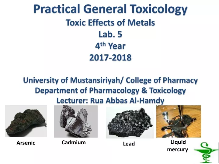
Practical General Toxicology Toxic Effects of Metals Lab. 5 4 th Year 2017-2018 University of Mustansiriyah/ College of Pharmacy Department of Pharmacology & Toxicology Lecturer: Rua Abbas Al-Hamdy. Liquid mercury. Cadmium. Arsenic. Lead. Lab objectives:

E N D
Practical General ToxicologyToxic Effects of MetalsLab. 54thYear2017-2018University of Mustansiriyah/ College of PharmacyDepartment of Pharmacology & ToxicologyLecturer: Rua Abbas Al-Hamdy Liquid mercury Cadmium Arsenic Lead
Lab objectives: • Objectives of this lab are to determine: • some toxic effects associated with arsenic (As), cadmium (Cd), lead (Pb), & mercury (Hg) which are major toxic metals. • biomarkers of metal exposure.
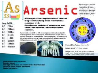
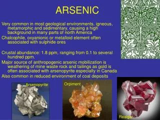
Arsenic: • Sources of exposure: • Environmental arsenic exposure mainly occurs from arsenic-contaminated drinking water. • Manufacture of pesticides, & herbicides. • Smelting industries.
Arsenic poisoning from groundwater in West Bengal: • In India arsenic contamination in ground water was first • reported in West Bengal in 1978.
Some effects of arsenic poisoning: • Acute poisoning: • Hair loss • Transverse bands of opacity in nails (Mees’ lines) (Fig 1) • Fatty degeneration of liver • Chronic poisoning: • Melanosis (neck, eyelids) • Hyperkeratosis (Fig 2) • Hyperpigmentation (rain drop pattern) (Fig 3) • Skin cancer (Fig 4)
Cadmium : • Sources of exposure: • Food due to the use of cadmium-containing water for irrigation of plants. • Cigarette smoking.
Some toxic effects of cadmium: • kidney damage. • Skeletal damage: Long-term high cadmium exposure may cause skeletal damage, first reported from Japan, where the itai-itai (ouch-ouch) disease (Fig 5) (a combination complications of osteomalacia & osteoporosis) was discovered in the 1950s. The exposure was caused by cadmium-contaminated water used for irrigation of local rice fields. • Prostate cancer & kidney cancer.
Figure 5. Itai-itai disease Rice fields
Source of exposure: • Young children are particularly vulnerable to the toxic effects of lead. • Environmental sources of lead exposure in children are shown in (Fig 6). • A major route of exposure for the general population is from food & water. • Other potential sources of lead exposure are battery making, soldering, jewelry making, & pottery making.
Some effects of lead: • The majority of lead which is absorbed is stored in the bones and teeth. In children, about 70% of lead is distributed in this way; in adults up to 95%. The hypermineralisation is reflected in the form of densities which are the classic “lead lines” observed on x-ray (Fig 7).
Figure 7. Longbone radiograph of knees - metaphyseal “lead band".
Microcytic & hypochromicanemia, as in iron deficiency. (Fig 8) Normal Hypochromic microcytic anemia Figure 8. Normal blood smear and a smear from a patient with hypochromic microcytic anemia.
Blood film examination may reveal basophilic stippling of red blood cells(dots in red blood cells visible through a microscope) (Fig 9). Figure 9. Basophilic stippled cell (arrowed)
Chronic lead nephrotoxicity: consists of interstitial fibrosis & progressive nephron loss, azotaemia & renal failure. • A remarkable pathogenic feature of lead poisoning is the presence of inclusion bodies composed of lead-protein complex (Fig 10). Lead-induced inclusion bodies are frequently nuclear, & are common in kidney.
Figure 10. Lead-induced inclusion body formation in kidneys from wild-type (WT) mouse. Arrow indicates typical karyomegaly of P3 proximal tubular cell.
In severe toxicity [Blood lead (BL) more than 100 mcg/100 ml], lead results in: • lead palsy: wrist drop (Fig 11) or foot drop. • a bluish black lead line on gums (Burton’s line) (Fig 12). • lead encephalopathy: It is more common in children.
Mercury: • Sources of exposure: • Breaking of mercury fluorescent light bulbs. • Liquid mercury following breakage of thermometers. • Dental amalgam. • Methyl mercury from consumption of fish.
Minimata disease: • Between 1953 & 1970, around Minamata Bay in Japan, more than 2000 people were diagnosed to be suffering from a curious cluster of neurological symptoms comprising paraesthesiae, narrowing of vision, dysarthria, diminution of hearing, amnesia, ataxia, staggering gait, weakness, & emotional instability. • Some developed paralysis & became stuporous, & out of all the people afflicted nearly a hundred died. The condition has been known as the Minimata disease (Fig 13).
It was caused by consumption of fish contaminated with methyl mercury. The most severely affected victims were actually infants who had been exposed in utero. Figure 13 . Minamata disease
Iraq grain disaster: The shocking tragedy in Iraq in 1971–72, when 500 people died out of a total of 6530 victims due to consumption of imported wheat and barley meant for sowing, treated with methyl mercury fungicide.
Some effects of mercury poisoning: • Acute poisoning from inhalation: • Dyspnoea, cough, fever, stomatitis • Deep red oral mucosa with “strawberry tongue” (Fig 14) • Skin rash (Fig 15)
Chronic poisoning by ingestion: • Colitis • Dementia • Tremor • Renal failure • Acrodynia (Pink disease) (Fig 16). This is seen mainly in children. The hands & feet become puffy, pinkish, painful, paraesthetic, perspiring & peeling.
Biomarkers of metal exposure: • The biological half-life varies according to the metal as well as the organ or tissue. For example, the biological half-lives of cadmium in kidney & lead in bone are 20 to 30 years, whereas for some metals, such as arsenic or lithium, they are only a few hours to days. • Blood, urine, & hair are the most accessible tissues for measuring metal exposure. • Blood and urine concentrations usually, but not always, are reflective of more recent exposures.
Hair can be useful in assessing variations in exposure to metals over the period of its growth. Analyses can be performed on segments of the hair, so that metal content of the newest growth can be compared with past exposures.