Download
1 / 1
10 likes | 129 Views
How effective propolis is on preventing bee viruses. Brianna Breiland, Amy Puckett, Paige Salmon, Nicole Wienke 1University of Wisconsin-River Falls, WI. Introduction

E N D
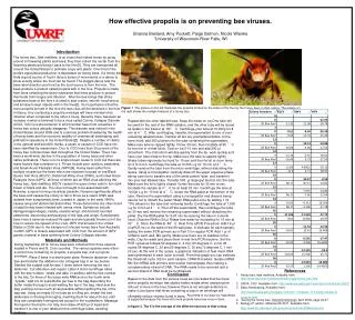
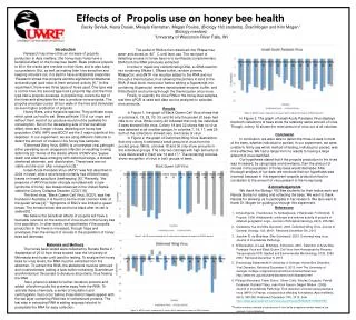
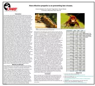
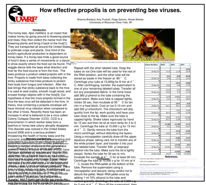
How effective propolis is on preventing bee viruses. Brianna Breiland, Amy Puckett, Paige Salmon, Nicole Wienke1University of Wisconsin-River Falls, WI Introduction The honey bee, Apis mellifera, is an insect that makes honey by going around to flowering plants and trees; they then collect the nectar from the flowering plants and bring it back to the hive[1]. They are transported all around the United States to pollinate crops and plants. One third of the world’s agricultural production is dependent on honey bees. If a honey bee finds a good source of food it does a series of movements or a dance to show exactly where the food can be found. The waggle dance tells the bees what direction and how far the food source is from the hive. The bees produce a product called propolis with in the hive. Propolis is made from bees collecting the sticky substance that trees produce to protect their buds from fungus and infection. After the bee brings that sticky substance back to the hive it is used to seal cracks, smooth rough wood, and encase foreign objects with in the hive[3]. Our hypothesis is that the more propolis formed in the hive the less virus will be detected in the hive. In theory, hive containing a propolis envelope will have minimal virus infection when compared to the other 2 hives. Recently there has been an increase in what is believed to be a virus called Colony Collapse Disorder (CCD). CCD is a phenomenon in which worker bees from a beehive or honey bee colony abruptly disappear. This disorder was noticed in the United States around 2006 and is a serious problem threatening the health of honey bees and the economic stability of commercial beekeeping and pollination operations in the United States[2]. Despite a number of claims in the general and scientific media, a cause or causes of CCD have not been identified by researchers. Due to CCD more than 30 percent of the honey bee colonies have died throughout the United States. There has been a world-wide decline in the population of honey bees and other native pollinators. There is not a single known cause to CCD but there are many factors that contribute to it. Those include poor nutrition, pesticides, and Israeli Acute Paralysis Virus (IAPV)[4]. Honey bees suffer from multiple viruses but the three which are research focused on are Black Queen Cell Virus (BQCV), Deformed Wing Virus (DWV), and Israeli Acute Paralysis Virus (IAPV), all three of which are an RNA virus affecting the honey bee, Apis mellifera. BQCV causes the queen larva cells to turn dark brown or black and die. The virus is thought to be associated with Nosema, a spore forming unicellular parasite. Nosema significantly affects the bees and causes the colony to fail to thrive as a result. DWV was isolated from symptomatic bees, located in Japan, in the early 1980’s, causes wing and abdominal deformities. These deformities are often found on adult honey bees infested with varroa mites. Symptoms include damaged appendages, stubby, useless wings, shorted and rounded abdomens, discoloring and paralysis of the legs and wings. Symptomatic bees have a severely-reduced life span and are typically thrown out of the hive to reduce the spread of the virus. IAPV first arrived in the United States in 2006 due to the transport of infected honey bees from Austrailia in 2005. IAPV is closely associated with CCD from the amount of IAPV genetic material in bees suffering from Colony Collapse disorder. . Figure 1. The picture on the left illustrates the propolis formed on the sides of the hive by the honey bees in their colony. The picture on the right shows the multiple features of a honey bee. Repeat with the other labeled tube. Keep the tubes on ice.One tube will be used for the rest of the RNA isolation, and the other tube will be stored as lysate in the freezer at -80 ° C. Centrifuge your tube at 10,000g for 8 min at 4 ° C. After centrifuging, transfer, the supernatant to one of your remaining labeled tubes. Transfer all but any precipitated debris. In the fume hood, add 380 µl phenol to the tube containing the supernatant. Make sure tube is capped tightly. Vortex 30 sec, then incubate at 95 ° C for ten min in a heat block. Cool on ice 5-10 min and add 200 µl chloroform. The chloroform will drip quickly from the tip- work quickly and have your tube close to the tip. Make sure the tube is capped tightly. Shake tubes vigorously by hand for 15 sec and then let sit at room temp for 2 to 3 min. Centrifuge the tube at 10,000 x g for 15 min at 4 ° C. Gently remove the tube from the micro-centrifuge, without disturbing the layers. Using a micropipettor carefully draw off the upper aqueous phase, taking care not to transfer any of the white protein layer, and transfer it into your last labeled tube. Transfer 500 µl isopropyl alcohol into the tube. Make sure the lid is tightly closed. Vortex the tube for 30 sec to mix. Incubate the sample at 4 ° C for at least 30 min. Centrifuge the tube at 10,000 x g for 10 min at 4 ° C, locate the RNA pellet at the bottom of the tube. Remove the supernatant using a micropipettor and discard, being careful not to disturb the pellet. Wash RNA pellet once by adding 1 ml 70% ethanol to the tube and vortexing briefly. Centrifuge the tube at 7,000 x g for 5 min at 4 ° C. Pour off the supernatant, then centrifuge the tube for 30 sec and remove the remaining supernatant without disturbing the pellet. Dry the RNA pellet for 5-20 min by opening the tube in a sterile hood. Dissolve RNA in 50 µl Rnase-free water by incubating for 10 min at 60 ° C. Store the RNA at -80°C. Real Time qPCR Procedure: Add 19 µl of qPCR mix to the wells of the 96-well plate, in triplicate for each sample, testing the same PCR primers as in Part 3 for regular PCR. Add 1 µl of cDNA to each well. Mix gently. Make sure there are no bubbles in the wells. Cap the wells and place them in real time PCR machine. Set the PCR cycles as follows:50 degrees C, 2 min; 95 degrees C, 2 min 45 cycles 95 degrees C, 20 sec;60 degrees C, 30 sec;72 degrees C, 1 min 20 sec. At the end of the cycles, a graphical indication of how much DNA was synthesized in each cycle is made. From that graph you can estimate the threshold cycle (Ct) for each sample. cDNA Procedure: Isolate mRNA. Mix the mRNA with primers and reverse transcriptase thus making a complementary strand of DNA. The RNA needs to be removed and a second strand of DNA must be synthesized. Based on the data from the various hives we concluded that the hives with a propolis envelope has slightly better results when analyzing the amount of virus in the hive, however there is not enough evidence to make it conclusive that hives containing a trap or an envelope will ultimately reduce viruses found in bees. According to our data our hypothesis is supported because the hives with more propolis have less virus in them. In figure 2. The X is the average of the different sections of that colony. Materials and Methods During September 2012, honey bees were collected from three apiaries located in Pierce and St. Croix counties. The various apiaries were either a control hive containing no propolis, a propolis trap, and a propolis envelope. Place 5 bees in a sterile petri plate. Remove abdomen of one bee and transfer the abdomen into a Kaypak bag in an ice bucket. Sterilize the scalpel and forceps 3 times before removing the next abdomen. Cut abdomen and repeat. Label 4 micro-centrifuge tubes with the bee number, initials and date. In addition add the bee number to the lids. On three of the tubes write RNA and on the fourth write “Lysate.” Add 0.5 ml Lysis Buffer per bee to the bag dispensing the buffer inside the bag to avoid wetting the top of the bag. Heat seal the bag, pushing out as much air as possible without getting the top of the bag wet. Using an empty 50 ml tube as a rolling pin, smash the bee abdomens in the bag thoroughly, mashing them for about 30 sec until they are completely homogenized except for the exoskeleton. Massage the liquid in the bad to mix fully, then draw off 620 µl of liquid and transfer it to one of your labeled micro-centrifuge tubes, avoiding bubbles. • References • Honey bee, Apis mellifera. Available from: http://www.fcps.edu/islandcreekes/ecology/honey_bee.htm • USDA: CCD. Available from: http://www.ars.usda.gov/news/docs.htm?docid=15572 • Basic Beekeeping. Available from:http://basicbeekeeping.blogspot.com/2011/12/lesson-113-sticky-subject-of-propolis.html • Saving the honey bee, Scientific American, April 2009, page 40-47 • Biology 9th edition, McGraw Hill,2011. Avaliable from: http://www.bio.davidson.edu/genomics/method/cDNAproduction Conclusion