Download
1 / 39
430 likes | 1.69k Views
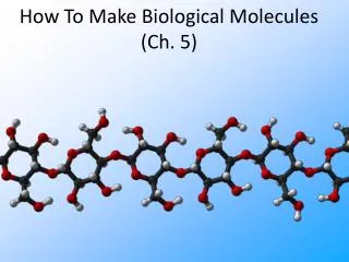
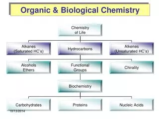
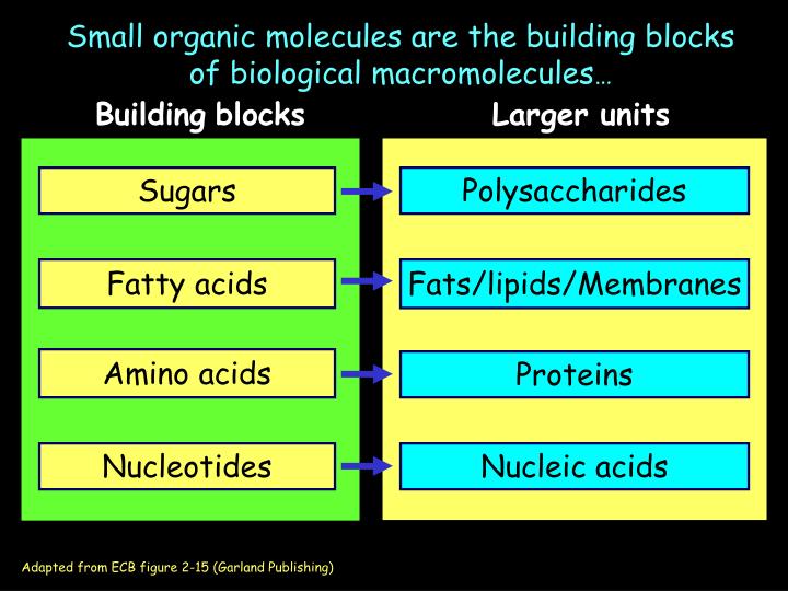
Sugars. Polysaccharides. Fatty acids. Fats/lipids/Membranes. Amino acids. Proteins. Nucleotides. Nucleic acids. Small organic molecules are the building blocks of biological macromolecules …. Building blocks. Larger units. Adapted from ECB figure 2-15 (Garland Publishing).

E N D
Sugars Polysaccharides Fatty acids Fats/lipids/Membranes Amino acids Proteins Nucleotides Nucleic acids Small organic molecules are the building blocks of biological macromolecules… Buildingblocks Largerunits Adapted from ECB figure 2-15 (Garland Publishing)
Plasmamembrane Various organelle membranes Most lipids are in membranes
Hydrophilic Carboxylic acid head group Hydrophobic hydrocarbon tail Fatty Acids (Amphipathic)
Stearic acid 18 carbons Saturated = no double bonds Oleic Acid (common in fats) 18 carbons Unsaturated 1 double bond, (common in oils) Fatty acids are distinguished by chain length and double bonds
Most lipids in cellsare formed by covalent bonds between fatty acids and glycerol ECB Fig. 11-10 Triacyl Glycerol = 3 fatty acids bonded to glycerol Ester bond - carboxylic acid and alcohol (animal fat, plant oils) Energy storage Very Hydrophobic
Polar Head Group = phosphate + polar moiety (Variable) glycerol Hydrophobic tails = fatty acid side chains Phospholipid - 3 partsMajor component of membranes
Phospholipid Hydrophilic region PhospholipidBilayer Hydrophobictail region
Lipid bilayer forms sphere in aqueous solution Forms barrier defining inside and outside spaces
Outer leaflet Lipids Inner leaflet Protein Cell Membrane-more complex Contains a variety of lipids, proteins, and carbohydrates Lipid bilayer 5 nm Multiple types of lipids are found in membranes Cytosol (inside) ECB Fig. 11-4
Glycolipids (sugar lipid) Phospholipids Sterols (cholesterol) serine Three Types of Membrane Lipid Moleculesall amphipathic ECB 11-7 galactocerebroside phosphatidylserine
Lecture 4 Membranes Fatty Acids Phospholipids Lipid bilayer Other membrane lipids Membrane properties Proteins Amino Acids Peptide bond Protein Structure
less ordered ordered Saturated straight hydrocarbon chains (no double bonds) Unsaturated hydrocarbon chains (with double bonds) Influence of FA saturation on lipid bilayer order Less ordered state increases membrane fluidity
Gel state Movement is greatly restricted (crystalline gel) Liquid state Hydrophobic tails free to move Membrane Fluidity (viscosity) Describes the physical state of the membrane Pure lipid bilayer - two states Transition temperature Liquid at temperatures Above the transition temp. Crystalline gel at temperatures below the transition temp Living cells require a fluid membrane, but not too fluid: Membrane fluidity is regulated by the cell 11.2-membrane_fluidity.mov
Melting points of 18-carbon Fatty Acids Fatty Acid Double bonds Melting point (˚C) Stearic acid 0 70 Oleic acid 1 13 2 -9 a-Linoleic acid Linolenic acid 3 -17 Membrane fluidity is governed by FA length and saturation 1. Fatty acid length - shorter the FA, the lower the transition temperature (melting point), favors liquid state 2. Fatty acid saturation - the more saturated, the higher the transition temperature, favors gel state 3. Presence of cholesterol - broadens the temperature over which transition occurs.
Polar head group Rigid Planar Steroid ring Nonpolar hydro- carbon tail Polar head group Stiffened region Fluid region Cholesterol stiffens lipid bilayers ECB 11-16 Mainly in animal cells, Not in plants
Glycolipids in outer leaflet Phospholipids Lipid composition varies in inner and outer leaflet
1. Spin (fast) 2. Lateral movement (less fast) 3. Flip-flop Almost never
Small hydrophobic Molecules O2, CO2, N2, benzene Lipid Bilayer Permeability Small Uncharged polar molecules H2O, glycerol, ethanol Large, uncharged Polar molecules Amino acids, glucose, nucleotides IONS H+, Na+, HCO3-, K+, Ca2+, Cl-, Mg2+ ECB Fig.12-2
Cell membrane ECB Fig. 11-4 Have discussed lipid bilayer, cholesterol, glycolipid Now move on to proteins
Sugars Polysaccharides Fatty acids Fats/lipids/Membranes Amino acids Proteins Nucleotides Nucleic acids Small organic molecules are the building blocks of biological macromolecules… Buildingblocks Largerunits Adapted from ECB figure 2-15 (Garland Publishing)
Proteins serve many functions in cells Transport proteins - move molecules across membranes Enzymes Structural proteins Motor proteins Signaling proteins Gene regulatory proteins Etc.
Amino Acids - the building blocks of proteins 20 different amino acids All amino acids have the same backbone, but the “R” group varies. See ECB Fig. 2-21
Polar Amino Acids Amino Acid Groups Based on chemical characteristics of R groups 1. Polar and negative charge (aspartic acid and glutamic acid) 2. Polar and positive charge (arginine, lysine, histidine) 3. Polar and uncharged (asparagine, glutamine, serine, threonine, tyrosine) 4. Nonpolar (alanine, glycine, valine, leucine, isoleucine, proline, phenylalanine, methionine, tryptophan, cysteine)
Negative charge Aspartic Acid (Asp, D) Glutamic Acid (Glu, E) Positive charge Histidine (His, H) Lysine (Lys, K) Arginine (Arg, R) Polar Charged Amino Acids (5)
Serine (Ser, S) Threonine (Thr, T) Asparagine (Asn, N) Glutamine (Gln, Q) Tyrosine (Tyr, Y) Polar Uncharged Amino Acids (5)
Non-polar amino acids (10 total) Alanine (Ala, A) Valine (Val, V) Leucine (Leu, L) Tryptophan (Trp, W) Phenylalanine (Phe, F) Methionine (Met, M) Isoleucine (Ile, I)
Glycine (Gly, G) Proline (Pro, P) Cysteine (Cys, C) Non-polar amino acids (cont’d)
+ H2O Peptide bond Polymerization of Amino Acids to Proteins Condensation rx Carboxyl end Amino end Dipeptide See also ECB figure 5-1
Lecture 4 Membranes Fatty Acids Phospholipids Lipid bilayer Other membrane lipids Membrane properties Proteins Amino Acids Peptidebond Protein Structure
1˚ structure: the linear sequence of amino acids N-terminal to C-terminal 4 Levels of Protein Structure 2˚ structure: stretches of the polypeptide chain that fold into a-helix or b-sheet (H-bonding) 3˚ structure: 3-dimensional conformation of a polypeptide chain 4˚ structure: multiple polypeptide chains interacting to form a complex
1˚ structure = sequence of amino acids (ECB Fig. 4-2)
Tertiary structure quaternary structure Secondary structure Higher levels of organization are determined by protein folding
Improper protein folding is associated with disease Prion diseases - scrapie (sheep), mad cow (bovine), chronic wasting disease (deer, elk), Creutzfeldt-Jacob disease (CJD, humans) Alzheimers and Huntingtons diseases - aggregated proteins in brain
Secondary Structure a-helix and b-pleated sheet
H bond C O H N helix R groups are on outside of helix H bond between peptide bonds, 4 a.a. apart
H bond pleated sheet
Disulfide bond formation (between cysteine residues) Tertiary Structure 3-D conformation of a singlepolypeptide chain Driven by many types of bonds (H-bonds, hydrophobic interactions, van der Waals, etc.)
Folding into tertiary structure forms domains in polypeptide Two different domains Single domain Polypeptide made up of several domains
Disulfide bridge Quaternary structure Multiple polypeptides interact via noncovalent and covalent (disulfide) bonds tetramer dimer