Download

1 / 9
140 likes | 1.51k Views
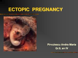
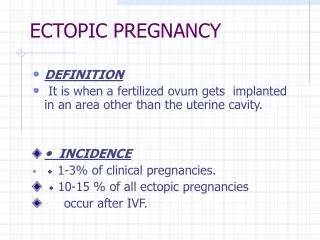
ECTOPIC PREGNANCY. is implantation of the fertilized ovum in any site other than the normal uterine location. Incidence: 1% of pregnancies. In 90% of these cases fallopian tubes other sites: ovaries, abdominal cavity

E N D
ECTOPIC PREGNANCY • is implantation of the fertilized ovum in any site other than the normal uterine location. • Incidence: 1% of pregnancies. • In 90% of these cases fallopian tubes • other sites: ovaries, abdominal cavity • Predisposing factors: tubal obstruction (50%), either due to chronic inflammatory changes in the oviduct; tumors; and endometriosis; IUCD.. • In 50% of tubal pregnancies, no anatomic cause can be demonstrated. • Early: normal early development of the embryo, with the formation of placental tissue, the amniotic sac, and decidual changes • Later: the placenta eventually burrows through the wall, causing intratubal hematoma (hematosalpinx), intraperitoneal hemorrhage. • Rupture of an ectopic pregnancy may be catastrophic, with the sudden onset of intense abdominal pain and signs of an acute abdomen, often followed by shock. Prompt surgical intervention is necessary.
Hydatidiform Mole: Complete and Partial • Are 2 forms of abnormal gestational processes. The two patterns result from abnormal fertilization • in a complete mole an empty egg is fertilized by two spermatozoa (or a diploid sperm), yielding a diploid karyotype composed of entirely paternal genes • In a partial mole a normal egg is fertilized by two spermatozoa (or a diploid sperm), resulting in a triploid karyotype with a predominance of paternal genes • Morphology: swollen cystically dilated chorionic villi (grapelike structures). The swollen villi are covered by varying amounts of mildly to highly atypical chorionic epithelium. • The complete hydatidiform mole does not permit embryogenesis and therefore never contains fetal parts. All of the chorionic villi are abnormal, and the chorionic epithelial cells are diploid (46,XX or, uncommonly, 46,XY). • The partial hydatidiform mole is compatible with early embryo formation and therefore contains fetal parts, has some normal chorionic villi, and is almost always triploid (e.g., 69,XXY).
incidence of complete moles 1 to 1.5 per 2000 pregnancies in the US and other Western countries. • For unknown reasons there is a much higher incidence in Asian countries. • Moles are most common before age 20 years and after age 40 years • a history of the condition increases the risk in subsequent pregnancies. • Early monitoring of pregnancies by ultrasound early diagnosis of hydatidiform mole. • Clinically: Elevations of hCG in the maternal blood and absence of fetal parts or fetal heart sounds are typical. • Prognosis: • complete moles: • 80% to 90% no recurrence after curettage • 10% invasive (invades myometrium) • 2% to 3% choriocarcinoma. • Partial moles: • better prognosis and rarely give rise to choriocarcinomas.
Choriocarcinoma • This very aggressive malignant tumor arises either from gestational chorionic epithelium or, less frequently, from totipotential cells within the gonads. • Choriocarcinomas are rare in the Western hemisphere, and in the United States they occur in about 1 in 30,000 pregnancies. They are much more common in Asian and African countries, reaching a frequency of 1 in 2000 pregnancies. • The risk is somewhat greater before age 20 and is significantly elevated after age 40. • 50% of choriocarcinomas arise in complete hyaditidiform moles; 25% arise after an abortion, and most of rest in normal pregnancy. • Stated in another way, the more abnormal the conception, the greater is the risk of developing gestational choriocarcinoma. • Clinically: appearance of a bloody, brownish discharge accompanied by a very high titer of hCG, in blood and urine, and the absence of marked uterine enlargement, such as would be anticipated with a mole.
Choriocarcinoma- Morphology • Grossly: very hemorrhagic, necrotic masses within the myometriumand into vessels of uterus. • Sometime, the primary lesion may self-destruct, and only the metastases are present. • Microscopically: In contrast to hydatidiformmoles and invasive moles, chorionic villi are not formed; instead, the tumor is purely epithelial, composed of anaplasticcuboidalcytotrophoblast and syncytiotrophoblast.
Prognosis • By the time most choriocarcinomas are discovered, there is usually widespread dissemination via the blood, most often to the lungs (50%), vagina (30% to 40%), brain, liver, and kidneys. • Lymphatic invasion is uncommon • Despite the extreme aggressiveness of these neoplasms, which made them nearly uniformly fatal in the past, current chemotherapy has achieved remarkable results. Nearly 100% of cases can be cured, even with some metastatic neoplasms to vagina and the lungs. • By contrast, there is relatively poor response to chemotherapy in choriocarcinomas that arise in the gonads (ovary or testis).