Download
1 / 27
280 likes | 550 Views
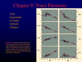
TRACE ELEMENTS IRON. IRON METABOLISM. DISTRIBUTION OF IRON IN THE BODY Between 50 to 70 mmol (3 to 4 g) of iron are distributed between body compartments.

E N D
IRON METABOLISM • DISTRIBUTION OF IRON IN THE BODY • Between 50 to 70 mmol (3 to 4 g) of iron are distributed between body compartments. • In normal subjects it is all protein-bound; in plasma it is bound to Transferrin, in the stores to protein in ferritin and haemosiderin, and in erythrocytes it is incorporated into hemoglobin.
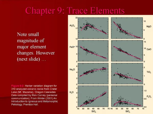
Distribution of Iron in a 70 kg Adult Male In an adult female of similar weight, the amount in stores would be generally be less (100-400 mg) and the losses would be greater (1.5 - 2 mg/d).
IRON METABOLISM • About 70 per cent of the total iron is circulating in erythrocyte hemoglobin. • Up to 25 per cent of the body iron is stored in the reticuloendothelial system, in the liver, spleen and bone marrow; bone marrow iron is drawn on for hemoglobin synthesis. Iron is stored as protein complexes, ferritin and haemosiderin. Ferritin iron is more easily released from protein than that in haemosiderin. Haemosiderin, probably an aggregate of ferritin, can be seen by light microscopy in unstained tissue preparations.
IRON ABSORPTION • The control of body iron content depends upon control of absorption by an active process in the upper small intestine. Within the intestinal cell some of the iron combines with the protein apoferritin to form ferritin, which, as elsewhere in the body, is a storage compound.
IRON ABSORPTION • Normally about 18 umol (1 mg) of iron is absorbed each day and this just replaces loss. This amounts to about 10 per cent of that taken in the diet, • Iron absorption seems to be influenced by any or all of the following factors: • oxygen tension in the intestinal cells; • marrow erythropoietic activity; • the size of the body iron stores.
IRON ABSORPTION • Iron absorption is also increased in many non-iron deficiency anaemias. • Most normal women taking an adequate diet probably absorb slightly more iron than men and so replace their higher losses in menstrual blood and during pregnancy. • Iron requirements for growth during childhood and adolescence are similar to, or slightly higher than, those of menstruating women and can be met by increased absorption from a normal diet.
IRON TRANSPORT IN PLASMA • Iron is transported in the plasma in the ferric form, attached to the specific binding protein, transferrin(2molecules/transferrin), at a concentration of about 18 umol/L (100 mg/dl). • Transferrin is normally capable of binding about 54 umol/L (300 mg/dl) of iron and is therefore about a third saturated. • Transferrin-bound iron is carried to stores and to bone marrow cells and in the latter some iron passes directly into developing erythrocytes to form hemoglobin.
Factors Affecting Plasma IronConcentration • Sex and age differences • Pregnancy and oral contraceptives • Variation within in individual • Random variation • Circadian (diurnal ) rhythm • Monthly variation in women
IRON EXCRETION • There is probably no control of iron excretion; loss from the body may depend on the ferritin iron content of cells lost by desquamation, mostly into the intestinal tract and from the skin. The total daily loss by these routes is about 18 umol (1 mg). Urinary loss is negligible, reflecting the fact that all circulating iron is protein-bound.
COMPARISON OF IRON LOSSES IN MEN AND MENSTRUATING/ PREGNANT WOMEN
PATHOLOGICAL FACTORS AFFECTING PLASMA IRON CONCENTRATION Iron deficiency and iron overload usually cause low and high plasma iron concentrations respectively. • Iron deficiency is associated with a hypochromic, microcytic anemia and with reduced amounts of stainable bone marrow iron. Plasma ferritin concentrations are usually, but not always, low. • Iron overload is associated with increased amounts of stainable iron in liver biopsy specimens and plasma ferritin conc are high.
PATHOLOGICAL FACTORSAFFECTING PLASMA IRONCONCENTRATION Other pathological factors • Any acute or chronic illness, even a bad cold • Disorders in which the marrow cannot use iron, either because it is hypoplastic, or because some other essential erythropoietic factor, such as vitamin B12 or folate, is deficient; • Hemolytic anemia. • Acute liver disease.
Transferrin and Total Iron-binding Capacity (TIBC) • Plasma iron concentrations alone give no information about the state of iron stores. • Diagnostic precision may sometimes be improved by measuring both the plasma transferrin and iron concentrations. • The total iron-binding capacity (TIBC). Is usually a valid measure of the transferrin concentration.
PHYSIOLOGICAL CHANGES IN THE PLASMA TRANSFERRIN CONCENTRATION • The plasma transferrin concentration is less labile than that of iron. However, it rises: • After about the 28th week of pregnancy even if iron stores are normal; • In women taking some oral contraceptive preparations; • In any patient treated with estrogens.
THE PLASMA TRANSFERRIN PATHOLOGICAL CHANGES IN CONCENTRATION Plasma transferrin concentration and TIBC: • Rise in iron deficiency and fall in iron overload • Fall in those chronic illnesses associated with low plasma iron concentrations • are unchanged in acute illness • May be very low in the nephrotic syndrome
SYNDROMES OF IRON OVERLOAD • IDIOPATHIC HAEMOCHROMATOSIS • ANAEMIA AND IRON OVERLOAD • DIETARY IRON OVERLOAD • INAPPROPIATE ORAL THERAPY
IRON OVERLOAD • CAUSES OF IRON OVERLOAD • Increased intestinal absorption: • idiopathic haemochromatosis; • anemia with increased, but ineffective, erythropoiesis; • liver disease (rare cause); • dietary excess; • inappropriate oral therapy.
CONSEQUENCES OF IRONOVERLOAD • Parenchymal iron overload occurs in idiopathic haemochromatosis and in patients with ineffective erythropoiesis. Iron accumulates in the parenchymal cells of the liver, pancreas, heart and other organs resulting in impairment of these organs & diabetes mellitus, hepatic carcinoma. • Reticuloendothelial iron overload is seen after excessive parenteral administration of iron or multiple blood transfusions. The iron accumulates initially in the R.E cells of the liver, spleen and bone marrow.
CONSEQUENCES OF IRONOVERLOAD • Haemosiderosis is a histological definition. An increase in iron stores as haemosiderin can be seen. It does not necessarily mean that there is an increase in total body iron; for example, in many types of anemia there is reduced hemoglobin iron (less hemoglobin) but increased storage iron. • Haemochromatosis describes the clinical disorder due to parenchymal iron-induced damage.
TRACE ELEMENTS CHROMIUM