Download
1 / 37
380 likes | 572 Views
Chapter 27 & 28. Prokaryotes & Protists. Ch. 27 ~ Prokaryotes. Overview: They’re (Almost) Everywhere! Most prokaryotes are microscopic But what they lack in size they more than make up for in numbers The number of prokaryotes in a single handful of fertile soil

E N D
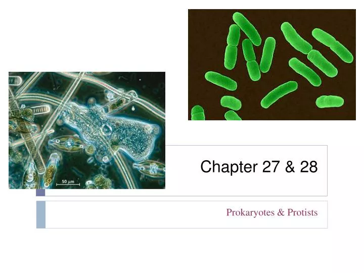
Chapter 27 & 28 Prokaryotes & Protists
Ch. 27 ~ Prokaryotes • Overview: They’re (Almost) Everywhere! • Most prokaryotes are microscopic • But what they lack in size they more than make up for in numbers • The number of prokaryotes in a single handful of fertile soil • Is greater than the number of people who have ever lived
Figure 27.1 • Prokaryotes thrive almost everywhere • Including places too acidic, too salty, too cold, or too hot for most other organisms
2 m 1 m 5 m (a) Spherical (cocci) (c) Spiral (b) Rod-shaped (bacilli) 27.1 ~ Structural, functional, and genetic adaptations contribute to prokaryotic success • Most prokaryotes are unicellular • Although some species form colonies • Prokaryotic cells have a variety of shapes • The three most common of which are spheres (cocci), rods (bacilli), and spirals
Cell-Surface Structures • One of the most important features of nearly all prokaryotic cells • Is their cell wall, which maintains cell shape, provides physical protection, and prevents the cell from bursting in a hypotonic environment
Lipopolysaccharide Outer membrane Peptidoglycan layer Cell wall Cell wall Peptidoglycan layer Plasma membrane Plasma membrane Protein Protein Gram- positive bacteria Gram- negative bacteria (a) Gram-positive. Gram-positive bacteria have a cell wall with a large amount of peptidoglycan that traps the violet dye in the cytoplasm. The alcohol rinse does not remove the violet dye, which masks the added red dye. 20 m (b) Gram-negative. Gram-negative bacteria have less peptidoglycan, and it is located in a layer between the plasma membrane and an outer membrane. The violet dye is easily rinsed from the cytoplasm, and the cell appears pink or red after the red dye is added. Figure 27.3a, b • Using a technique called the Gram stain • Scientists can classify many bacterial species into two groups based on cell wall composition, Gram-positive and Gram-negative
200 nm Capsule Figure 27.4 • The cell wall of many prokaryotes • Is covered by a capsule, a sticky layer of polysaccharide or protein
Fimbriae 200 nm Figure 27.5 • Some prokaryotes have fimbriae and pili • Which allow them to stick to their substrate or other individuals in a colony
Flagellum Filament 50 nm Hook Cell wall Basal apparatus Plasma membrane Figure 27.6 Motility • Most motile bacteria propel themselves by flagella • Which are structurally and functionally different from eukaryotic flagella
0.2 m 1 m Respiratory membrane Thylakoid membranes (a) Aerobic prokaryote (b) Photosynthetic prokaryote Figure 27.7a, b Internal and Genomic Organization • Prokaryotic cells • Usually lack complex compartmentalization • Some prokaryotes • Do have specialized membranes that perform metabolic functions
Chromosome 1 m Figure 27.8 • The typical prokaryotic genome • Is a ring of DNA that is not surrounded by a membrane and that is located in a nucleoid region
Endospore 0.3 m Figure 27.9 • Some species of bacteria • Also have smaller rings of DNA called plasmids • Draw a prokaryotic cell and label the DNA and plasmids • Prokaryotes reproduce quickly by binary fission • And can divide every 1–3 hours • Many prokaryotes form endospores • Which can remain viable in harsh conditions for centuries
27.2 • A great diversity of nutritional and metabolic adaptations have evolved in prokaryotes • Examples of all four models of nutrition are found among prokaryotes: • List Energy source & Carbon source: • Photoautotrophy • Chemoautotrophy • Photoheterotrophy • Chemoheterotrophy
Major nutritional modes in prokaryotes Table 27.1
27.3 • Molecular systematics is illuminating prokaryotic phylogeny • Until the late 20th century • Systematists based prokaryotic taxonomy on phenotypic criteria • Applying molecular systematics to the investigation of prokaryotic phylogeny • Has produced dramatic results
Domain Archaea Domain Eukarya Domain Bacteria Proteobacteria Gram-positive bacteria Nanoarchaeotes Crenarchaeotes Euryarchaeotes Korarchaeotes Cyanobacteria Spirochetes Chlamydias Eukaryotes Gamma Epsilon Alpha Beta Delta Universal ancestor • A tentative phylogeny of some of the major taxa of prokaryotes based on molecular systematics Figure 27.12
Bacteria • Diverse nutritional types • Are scattered among the major groups of bacteria • The two largest groups are • The proteobacteria and the Gram-positive bacteria
Chlamydias, spirochetes, Gram-positive bacteria, and cyanobacteria 2.5 m Chlamydia (arrows) inside an animal cell (colorized TEM) 5 m Leptospira, a spirochete (colorized TEM) 1 m 5 m Hundreds of mycoplasmas covering a human fibroblast cell (colorized SEM) Streptomyces, the source of many antibiotics (colorized SEM) 50 m Two species of Oscillatoria, filamentous cyanobacteria (LM) Figure 27.13
Table 27.2 Archaea • Archaea share certain traits with bacteria • And other traits with eukaryotes
Some archaea live in extreme environments • Extreme thermophiles thrive in very hot environments • Extreme halophiles live in high saline environments Colorful “salt-loving” archae live in these ponds near San Fransisco. Used for commercial salt production. • Methanogens • Live in swamps and marshes and produce methane as a waste product
27.4 • Prokaryotes play crucial roles in the biosphere • Chemical Recycling • Prokaryotes play a major role in the continual recyclingof chemical elements between the living and nonliving components of the environment in ecosystems • Chemoheterotrophic prokaryotes function as decomposers • Breaking down corpses, dead vegetation, and waste products • Nitrogen-fixing prokaryotes • Add usable nitrogen to the environment
Figure 27.15 Symbiotic Relationships • Many prokaryotes - • Live with other organisms in symbiotic relationships such as mutualism and commensalism
5 µm Figure 27.16 27.5 • Prokaryotes have both harmful and beneficial impacts on humans • Some prokaryotes are human pathogens • But many others have positive interactions with humans • Prokaryotes cause about half of all human diseases • Lyme disease is an example
Prokaryotes in Research and Technology • Experiments using prokaryotes have led to important advances in DNA technology • Prokaryotes are the principal agents in bioremediation • The use of organisms to remove pollutants from the environment • Prokaryotes are also major tools in • Mining, the synthesis of vitamins, production of antibiotics, hormones, and other products
Ch. 28 ~ Protists • 28.1: Protists are an extremely diverse assortment of eukaryotes • Protists are more diverse than all other eukaryotes • And are no longer classified in a single kingdom • Most protists are unicellular, colonial or multicellular
Protists, the most nutritionally diverse of all eukaryotes, include • Photoautotrophs, which contain chloroplasts • Heterotrophs, which absorb organic molecules or ingest larger food particles • Mixotrophs, which combine photosynthesis and heterotrophic nutrition
There is now considerable evidence that much of protist diversity has its origins in endosymbiosis
28.3: Euglenozoans have flagella with a unique internal structure • Euglenozoa is a diverse clade that includes • Predatory heterotrophs, photosynthetic autotrophs, and pathogenic parasites
Dinoflagellates • Are a diverse group of aquatic photoautotrophs and heterotrophs • Are abundant components of both marine and freshwater phytoplankton • Ciliates, a large varied group of protists • Are named for their use of cilia to move and feed • Have large macronuclei and small micronuclei • Conjugation – Two cells exchange haploid micronuclei (similar to prokaryotic conjugation). • **Produces genetic variation
28.5 Hairy and smooth flagella1. Diatom – Surrounded by a two part glass like wall and are a major component of phytoplankton. Phytoplankton account for half of the photosynthetic activity on earth – responsible for much of the oxygen in our atmosphere.
2. Golden Algae – Have two flagella and their color results from carotenoids.
3. Brown Algae – multicellular mostly marine protists, commercially used; seaweeds
28.6 Threadlike pseudopodia • Pseudopodia extend and help to injest microorganisms.
28.7 ~ Amoebozoans Lobed shaped pseudopodia *Entamoebas – parasites of vertebrates and cause amebic dysentry in humans. *Slime molds – extend their pseudopodia through decomposing material, engulfing food by phagocytosis.
28.8 ~ Red & Green Algae are the closest relatives of land plants.