Download
1 / 10
110 likes | 1.26k Views
DIRECT REPAIR OF LUMBAR SPONDYLOLYSIS BY SEGMENTAL PEDICLE SCREW-INFRALAMINAR HOOK CONSTRUCT. Cagatay OZTURK, MD Ahmet ALANAY, MD Meric ENERCAN, MD Selhan KARADERELER, MD Omer KARATOPRAK, MD Azmi HAMZAOGLU, MD Istanbul Spine Center Florence Nightingale Hospital Istanbul-TURKEY. PURPOSE.

E N D
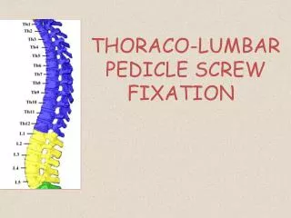
DIRECT REPAIR OF LUMBAR SPONDYLOLYSIS BY SEGMENTAL PEDICLE SCREW-INFRALAMINAR HOOK CONSTRUCT Cagatay OZTURK, MD Ahmet ALANAY, MD Meric ENERCAN, MD Selhan KARADERELER, MD Omer KARATOPRAK, MD Azmi HAMZAOGLU, MD Istanbul Spine Center Florence Nightingale Hospital Istanbul-TURKEY
PURPOSE • To analyse the safety and efficacy of direct pars repair by using segmental pedicle screw-infralaminar hook construct. • Twenty patients (16 female and 4 male) who had treated by direct pars repair with segmental pedicular screw-hook fixation and with minimum 2 years follow-up were included in this study. PATIENT SAMPLE
METHODS • Allpatients had spondylolysiswithisthmicdefect at • L5 (n=18) • L4 (n=2) • Six (30%) of them had grade 1 spondylolysis. • 6 patients (30%) had mildscoliosis • 3 (15%) had Schuermannkyphosis. • Onepatient had bothscoliosisandScheuermankyphosis. • All had lowbackpainunresponsivetoconservativemeasuresfor at least 6 months. • None had radiculopathysigns.
METHODS • All patients had a preoperative CT scan and magnetic resonance imaging and all had Phirman class I healthy discs at the involved level. • All patients had CT scans at the postoperative 1 year follow-up to evaluate healing. • Two-year follow-up x-rays were analysed in terms of disc degeneration and collapse at the operated level and progression of existing deformities.
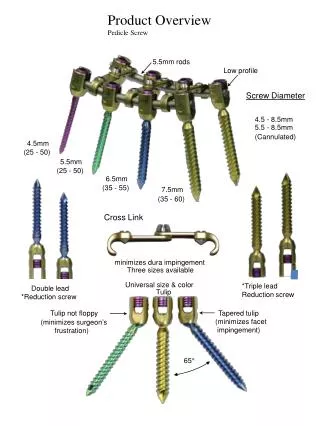
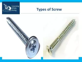
Hook-pedicle screw fixation(Tokuhashi technique) (1996) Placement of pedicle screws Removal of fibrous tissue Exposure of pars defect Onlay autograft harvested from posterior superior iliac spine Autograft placement on to the defect Placement of infralaminar hooks Rod fixation and compression
RK, 14y, F Right Left
RESULTS • The mean follow-up period was 34.8 (range; 24 to 72) months. • Mean age was 16.8 (range; 14 to 18) years. • CT scan revealed succesfull healing in all patients. • None of the patients had degenerative findings at the disc level below the pars defect.
CONCLUSION • Surgical treatment of adolescent patients with spondylolysis by using pedicle screw-infralaminar hook technique resulted with satisfactory clinical and radiological outcome.