Download
1 / 273
2.9k likes | 4.08k Views
Homograft, Xenograft & Bioprosthesis. Seoul National University Hospital Department of Thoracic & Cardiovascular Surgery. Valved Homografts. Introduction Valved homografts , introduced in the 1960s, have used for reconstruction of the right ventricular outflow tract .

E N D
Homograft, Xenograft& Bioprosthesis Seoul National University Hospital Department of Thoracic & Cardiovascular Surgery
Valved Homografts • Introduction • Valved homografts, introduced in the 1960s,have used for reconstruction of the right ventricular outflow tract . • Advantages of homografts, such as ease of handling and improved hemostasis, have been major contributing factors • Viable endothelial cells and perhaps also viable fibroblasts, thus contributing to an antigenically active cells, which could lead to a more intense host response. • This increased immunogenicity could perhaps be an important contributing factor for accelerated fibrocalcifications, particularly in very young patients.
Homograft • Processing • Cryopreservation • Antibiotics • Fetal calf serum • RPMI media • DMSO • Thawing • Harvest of aortic, mitral, pulmonary valve & others
Sterilization for Homograft • Antibiotics for uses • Modified Hanks solution or TCM • Cefoxitin sodium ( 240mg/L ) • Vancomycin hydrochloride ( 50mg/L) • Lincomycin hydrochloride ( 120mg/L) • Polymixin B sulfate ( 100mg/L) • Amphotericin B ( 25mg/L )
Homograft Processing • Essence • 1. Possible procurement on beating heart, but • non-beating heart within 24 hrs after death • 2. Reduce antibiotic incubation time to 6 hrs • 3. Fibroblast is important in the maintenance • of the valve matrix. • * Viable cells persist, produce collagen and • repair damaged matrix. • 4. Low dose antibiotics and no amphotericin B
Homograft • Laboratory evaluation • Hematoxylin-eosin & elastica van Gieson • Elastic fiber of vessel wall • Muscle actin positive cell • Immunohistochemical staining • Flow cytometry • Antibodies for inflammatory cell • CD3 for all T lymphocytes • CD4 for TH lymphocytes and macrophages • CD8 for TC lymphocytes and NK cells • Scanning EM for endothelial integrity
Homograft • Advantages 1. Technical ease of implantation because they are soft & mold easily with patient’s tissue, resulting in better hemostasis in complex operations 2. Better hemodynamics than porcine-valved dacron • conduit, which improves the RV function after • operation. • 3. The branch of the homograft may be used to patch • distal pulmonary artery stenoses
Homograft • Durability of aortic valve • Homograft can last 20 years, essentially as a dead piece of tissue(become acellular within a few months), free from any mechanical reinforcement by crosslinking agents. • Unknown exactly what features of the native valve tissue give it such remarkable durability than other bioprosthesis, the importance of its internal complexity is being appreciated more and more. • Presence of interconnected sheets of collagen, layers and tubes of elastin, highly nonlinear mechanics, anisotropy, and viscoelasticity endow the valve tissue with is unique longevity.
Cryopreservation • Methods • Treatment with antibiotic agents at 4 ℃ for 24 hours or culture medium with antibiotics • Frozen in tissue culture medium(TCM) 199 solution containing 10% calf serum, 10% DMSO, and 5% HEPES buffer, with a cooling velocity at a rate of -1 ℃ / min to -80 ℃ with programmable controlled-rate freezer. • Frozen xenograft is put into sealed package and preserved in vapor phase of liquid nitrogen at -196 ℃ • The cryopreserved xenograft is thawed in a water bath at 37 ℃ quickly
Cryopreservation • Protocol • Transport Maximum 24 h at 4°C RPMI nr. 1640 300 mL • Sterilization 14–18 h at 4°C RPMI nr. 1640 300 mL 24 mg Mefoxin. 12 mg Lincomycine 5 mg Vancomycine. 100000 UI Colymicine • Storage –150°C untilimplant 90 g Albumine 4% 10 mL DMSO DMSO = dymethylsulphoxide; RPMI = Roswell Park Memorial Institute.
Cryopreservation • Influence on immunity • Cryopreservation technique might attenuate allograft immunogenicity by reducing the viability and cellularity of the endothelium or by diminishing the expression of leukocyte adhesion molecule • Cryopreservation technique reduces the xenogeneic immunogenicity of endothelial cells as in allograft • Cryopreservation technique could not reduce the immunogenicity of other cellular & extracellular matrices of xenograft
Cryopreservation • Effects on cells • Cytosolic and mitochondrial functions of endothelial cells were damaged seriously during cryopreservation, especially in thawing process, and those process may cause a latent cytosolic and mitochondrial injury, even in the fibroblast • Viability of donor fibroblast which can remodel the matrix assembly is damaged by cryopreservation and thawing, that may cause collagenolytic activation and degradation of collagen synthesis in a cryopreserved valve, which will lead to destruction of matrix
Cryopreservation • Effects on valve tissue • The valvular extracellularmatrix(ECM ) contains a variety of structures, underliesand surrounds the interstitial cells, and performs many essentialfunctions, including mechanical support and physical strength • ECM exerts profound influenceson cell adherence, migration, and differentiation as well asthe pattern of gene expression of the cells in contact withit • The quality of the structural matrix at implantationmay predetermine durability or failure of a cryopreserved heartvalve (collagenous bundles and elastin-containingfibers) based on different techniques before implantation
Cryopreservation • Effects on valve matrix • Conventional approaches to cryopreservation ofheart valves have a detrimental and destructive effect on crucialleaflet matrix elements. • Serious alterationand significant deterioration of collagenous and elastic fiberstructures, accompanied by a general damage of the leaflet histoarchitecturecaused by extracellular ice formation. • Alternative preservationtechniques proposed for clinical use, such as vitrification, D-hydro-, or glycerol-treatment, are crucial.
Cryopreservation • Mechanismsof structural alteration • Related to anactivation of metalloproteinases (MMPs) upon thawing, anddisruption of normal extracellular matrix metabolism by thevalve interstitial cells, a function shown to be impairedin cryopreserved tissue • Side specific cell phenotypes anddifferences in extracellular matrix organization may accountfor the heterogeneity in extracellular matrix alteration. • The lack of crimp in the thawed cryopreservedvalves suggests that the biomechanical properties of the ventricularislayer have been altered
Cryopreserved Homografts • Examination parameters • Tissue structure • Tissue viability • Cell proliferative capacity • Metabolic function • Identification of cell-specific antigens ( Immunohistochemistry) • Flow cytometry( MHC I and II antibody)
CryopreservedHomograft • Viability test • 1. Radiolabeled thymidine • 2. Collagen synthesis • 3. Protein synthesis • 4. Dye uptake & dye exclusion • 5. Autoradiography
Homograft Viability Test • Methods for viability • Cytometry using propidium iodure and fluorescent diacetate (FDA). • Double staining with fluorescein diacetate-propidium iodure (FDA-PI) is reported to be a rapid method for assessing cell viability compared with the trypan blue dye exclusion method
Cryopreserved Homografts • Structural fate • Viable fibroblasts synthesize main constituents of extracellular matrix: collagen, elastin, reticulin, and mucopolysaccharides, therefore longevity of the implantation is likely related to the viability of fibroblasts in the implanted valves • However, fibroblast in the donor allograft were unable to survive long, because of apoptosis. • Endothelial cells exhibit strong antigenicity, but can not survive under ischemic conditions and moreover, during the cryopreservation process endothelial cells lose their ability to proliferate. • The homograft will lose its endothelial cells and fibroblasts , and eventually become a non-viable tissue.
Cryopreserved Homograft • Standard model • This demonstrates preservation of endothelial & valve architecture & viability • This viability may not be a positive attribute, as initial thought, it contributes to immunogenicity and elicits a more vigorous immunologic reaction from the recipient. • Heightened immune response contributes to accelerated degeneration of allograft valve • An immunologically neutral graft has therefore been considered desirable, leading to new development that is decellularized to avoid introduction of immunogenic cells into the recipient
Cryopreservation • Effects on immune reaction • There were donor-specific immune responses evoked by allograft insertion, and on the other hand , clinically explanted allografts showed no evidence of rejection • Cryopreservation causes the greatest delay & diminuation of the expression of leukocyte adhesion molecules • Cryopreservation attenuates the allograft immunogenicity by reducing the viability and cellularity of endothelium • But it seems to be impossible to reduce xenogeneic immunogenicity with cryopreservation because of the marked destruction of other cellular and extracellular components
Aortic Valve Replacement • Advantages of homograft • Good hemodynamics, • Fewer thromboembolic complications • Avoidance of anticoagulation • Suitability in the presence of infection • Disadvantages of homograft • Risk of early failuredue to technical error • Limited durability and availability
Homograft Implantation • Surgical indication of endocarditis • Hemodynamic instability • Microbiology resistance, usually involved in active aortic endocarditis including vegetations, cusp destruction, and periannular extensions. • Aortic vegetations, especially when their diameter is superior to 10 mm, may be responsible for systemic embolization • Anatomic lesions progression are traditional complications leading to early surgery • Aortic periannular extension can degenerate in abscesses, discontinuity, fistulas, aberrant communications and false aneurysm, increasing congestive heart failure, and mortality rate
Homografts Implantation • Aortic position • pulmonary autograft is not recommendedfor young rheumatic aortic valve patients, particularly withmitral valve involvement • Ross procedure could be performedin young aortic valve patients if their mitral and pulmonaryvalves are intact • HAVR should be tailored to individual patients soas to choose the scalloped subcoronary or root replacement • In view of the long-term prognosis, the surgeon should use,if possible, the subcoronary replacement technique, which makesthe reoperation much easier
Homografts Implantation • Pulmonary position • Superior conduit durabilityin Ross patients has emphasized the placement of the homograftin the orthotopic pulmonary position and less sternal compressionof anteriorly placed conduits • The predominant mechanismof homograft stenosis was a poorly understood inflammatory reaction based on their noting of early onset ofstenosis, rapid clinical progression • Stretchingand lengthening of the homograft causing release of tissue factorswas one possibility suggested and the extent of that phenomenon might relate to the degree ofperipheral vascular distortion present
Homograft Failure • Etiologic factors • Factors unrelated to immunologic injury would include mechanical factors, such as sternal compression, or oversizing or undersizing of conduits with anatomic distortion, in addition to ischemic injury. • Factors related to immunologic injury include ABO incompatibility and human leukocyte antigens (HLA) incompatibility • Tissue processing steps such as warm ischemic time, antibiotic disinfection, cryopreservation, and thawing all affect viability of the grafts • More recently, investigators have demonstrated significant cellular infiltration with T-cells and B-cells in failed explanted valved allografts in children
Homograft Failure • Specials in young • The most important factors are size of conduit & age at implantation. • Primary reason, growth of young child over time • Secondary reason, decrease in the functional lumen of the conduit due to calcification or immune related • Anatomic type of the conduit, aortic or pulmonary • Heightened immune reaction to the implanted tissue • There is evidence that children produce a virulent T-cell response, where adults mount a much weaker response
Homograft Failure • Risk Factors • 1. Younger recipient age • 2. Aortic homograft related to higher elastin • & intrinsic calcium content • 3. Small homograft valve size • 4. Long aortic cross-clamp time at operation • 5. Pulmonary hypertension • 6. Distal pulmonary artery disease • 7. Younger donor age ( increased immunogenecity ) • 8. Technical factors • 1) Hood extension of proximal suture line • 2) Anatomic versus nonanatomic placement • 3) Compression of conduit
Homograft Failure • Alternatives to homograft • After a period of 10 years, 30% of the children initiallycorrected with allografts and 70% of the patients with xenograftshad undergone replacement of their initial conduits. • We haveavoided using an allograft or heterograft conduit in most patientswith tetralogy of Fallot or pulmonary atresia with ventricularseptal defect. • We have preferred to construct a transannularpatch with an underlying PTFE monocusp • Untilrecently, cryopreserved allografts have been the best valvedconduits and the limited durabilityof the allograft conduit became evident within a few years
Implanted Allografts • Failure process • Transplanted cryopreserved homografts rapidly lose their cellular components during the first year of implantation while the normal tissue architecture is damaged. • The allografts are capable of eliciting a cellular and humoral immune response, it is not yet clear what exact role the immune response plays in ECM damage • After transplantation, indicators of immune-mediated injury as persistent up-regulation of leukocyte adhesion molecules, the presence of neutrophyl granulocytes, depositions of antibodies and complement are missing in early phase after implantation.
Implanted Allografts • Loss of cellularity • Immunologic & chemical injury • Allografts are eliciting a cellular & • humoral immune response, not yet clear • what exact role the immune response • Hypoxia during valve processing • Reperfusion injury at implantation
Homograft • Immune responses • Children & adults respond differently to homograft. • Younger donor age related to increased immunogenicity • There is evidence that children produce a virulent T-cell response, where adults mount a much weaker response. • In addition, laboratory studies have demonstrated that allograft valve destruction is T-cell mediated. • Other HLA loci and nonimmunologic factors, including growth, degeneration, and technique, play a role in homograft failure
Implanted Allografts • Immune responses • All allograft recipients are likely to form IgG- and T-cell-mediated reaction to donor HLA antigens • Donor-specific T cells are likely to be the main agents of allograft injury, effected through secretion of high levels of cytokines • The location of induction and amplication of the immune response to the allograft remains unknown. • Dendritic cells, which is identified in human great vessels, together with endothelial cells are capable of presenting foreign HLA class I and II antigens to recipient T cells
Implanted Allografts • Immune responses • Valve leaflet cellularity was demonstrated to be significantly reduced with time • Donor-specific immune activation, some early infiltration of immune effector cells, such as macrophages and T lymphocytes is demonstrated • Most of the evidence for immune-mediated damage in this animal model occurred in the first 2 weeks after implantation, and by 4 weeks the valves were largely acellular • This observation may in fact represent evidence of immune-related valve injury, despite the absence of up-regulation of cell adhesion molecules
Homograft Failure • Fate of implanted homograft • Virtually all cryopreserved homografts demonstrate acellularity within a year of implantation • The lack of cells and cellular function limits tissue growth, performance, and reparative capacity; such "tissues" typically scar and then mineralize, which often leads to dysfunction. • Chronic rejection of cells in classically cryopreserved allograft tissues promotes the migration of inflammatory cells, exacerbating tissue degeneration, fibrosis, and functional failure
Homograft Implantation • Reduce immune response • One would be to match all allografts by blood type and HLA type. • Another would be to somehow remove the immunogenicity of valved allograft tissue, perhaps by removing the antigen presenting cells on the allograft • The third option would be to immunosuppress the recipient in order to abrogate the immune response of the recipient to the donor allograft.
Blood Cells • Production • In children, blood cells are produced in the marrow of all bones. • After the age of 20 years, only the marrow of the vertebrae, sternum, and ribs remain significantly active (red marrow) in the production of erythrocytes, many leukocytes, and platelets. • Active marrow contains pluripotential stem cellsthat are capable of replacing bone marrow completely (self-renewal) or can be diverted from self-renewal toward separate pools of committed progenitor cells
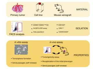
Stem Cells & Progenitor Cells • Characteristics • Progenitor cells : (1) they have lost the capacity for self renewal and (2) they are not pluripotential but are committed to produce (under the proper growth conditions) daughter cells of a particular type. During maturation, each cell line, promoted by a variety of stimulating factors, acquires distinctive properties • Pluripotent stem cell can differentiate in a few divisions into one of six classes of progenitor cells
Stem Cells & Progenitor Cells A pluripotent stem cell can differentiate in a few divisions into one of six classes of progenitor cells that go on to produce blast cells. Blast cells are the earliest morphologically distinct precursors of specific cell types. EPO = erythropoietin; GCSF= granulocyte colony-stimulating factor; GM-CSF = granulocyte-macrophage colony-stimulating factor; IL = interleukin; M-CSF = monocyte colony-stimulating factor; PMNs = polymorphonuclear monocytes.
Red Blood Cell • Immunologic properties • The red cells of different individuals differ to a very small extent in the structure of some carbohydrates that are part of membrane glycolipids • These differences bestow antigenic properties on red blood cells and cause red cell agglutination • ABO antigens • The A and B antigens are the most important of the more than 100 different blood group antigens that have been identified • In neonatal life, antibodies quickly develop against the antigens that are not present on our red cells, and these antibodies, called agglutinins, are carried in plasma. • The antigens are called agglutinogensand are carried on the red cells in the blood as well as on cells in many other tissues
Red Blood Cell • Rh antigens • Within the Rh system, the C, D, and E antigens are most important. They are found only in red cells. • D is the most antigenic component, and the presence or absence of D is designated as “Rh-positive” or “Rh-negative,” respectively. • 85 % of Caucasians & more than 99% of Asians are Rh+ • Anti-D develops only when the blood of a D– individual is exposed to D+ red cells. • This can occur as a result of transfusion or when Rh+ fetal blood mixes with the circulation of an Rh– mother
Antigens of ABO group A, H antigen that is present in individuals with type O blood. B, A antigen (type A blood) has a terminal N-acetylgalactosamine (NAG). C, B antigen (type B blood) has a terminal galactose (Gal). Cer = ceramide; Fuc = fucose; Gal = galatose; Glu = glucose.
Human Blood • Components of plasma • The major plasma proteins are albumin (4.5 g/dL), several globulins(2.5 g/dL), and fibrinogen(0.3 g/dL). • Most are synthesized by the liver, and they have five major functions: (1) carriers for hormones, trace metals, or drugs; (2) proteolytic agents in the cleavage of various hormonal or enzymatic precursors; (3) protease inhibitors; (4) source of plasma colloid osmotic pressure;(5) source of the humoral immunity portion of the immune system.
Arachidonic Acid Derivatives • Derivatives of arachidonic acid play a crucial role in platelet adhesion • to the endothelium, and the clotting process can be disrupted by • interruption of arachidonic acid metabolism.
Vascular Endothelium • Roles in hemostasis
Complement System • Natures • A system of 11 plasma enzymes identified as C1 to C9; C1 consists of the three subunits C1q, C1r, and C1s. The enzymes circulate in the inactive form but can be activated tolyse foreign cells. The activation proceeds in a step-like fashion, each activated enzyme hydrolyzing a peptide bond in the next inactive enzyme • Classical pathwayis triggered when immunoglobulin G or M binds to cell surface antigens • Alternate pathwayof complement activation does not require binding of immunoglobulins to cell surface antigens and triggered
Complement System • Activation • Classical pathway ; The consequent activation cascade eventually leads to (a) activated C3, a cell surface-associated factor that promotes opsonization, and (b) activated (C5, C6, C7, C8, C9), which is associated with production of chemotactic substances, release of histamine, and insertion of perforins into the plasma membrane. • Alternate pathway ; It is triggered when the circulating protein factor I attaches to specific surface polysaccharides in a bacterium or virus. This pathway also leads to activation of C3 and (C5, C6, C7, C8, C9) and their associated opsonization or cell lysis.
Monocytes • Function • Monocytes are formed in the bone marrow, enter the blood, and circulate for about 3 days before they enter the tissues by diapedesis and become tissue macrophages that differentiate to perform specific functions in different tissues. • They are phagocytic cells and perform many of the same actions that are performed by neutrophils • By secreting a large number of lysosomal, chemotactic, complement-activating, and pyrogenic factors, they are key effectors in the elimination of microorganisms and play an important role in immunity and blood clot formation.