Download
1 / 43
1.07k likes | 5.94k Views
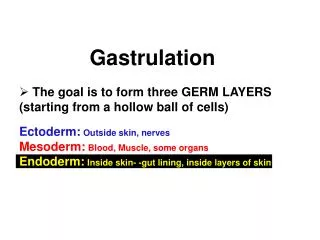
Today's Goals. Become familiar with the concepts of Cleavage, Gastrulation and Axis DeterminationBecome familiar with the types of cell movements in the embryoDescribe the processes of Cleavage and Gastrulation in Sea Urchin and Xenopus embryos. Sea Urchin Cleavage. Radial Holoblastic CleavageFi

E N D
1. Cleavage and Gastrulation - Sea Urchin and Frog Gilbert - Chapter 8 pp. 217-228
& 10 pp. 291 - 299
2. Today�s Goals Become familiar with the concepts of Cleavage, Gastrulation and Axis Determination
Become familiar with the types of cell movements in the embryo
Describe the processes of Cleavage and Gastrulation in Sea Urchin and Xenopus embryos
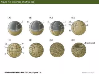
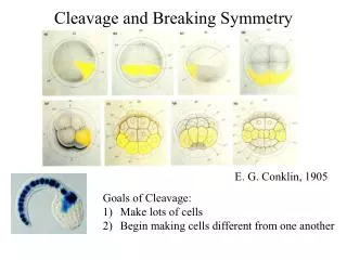
3. Sea Urchin Cleavage Radial Holoblastic Cleavage
First two divisions
Meridional, perpendicular to each other
Third division
Equitorial, perpendicular to first 2
Divides into animal half, vegetal half
5. Cleavage in Sea Urchins (cont.) Fourth cleavage
Animal half divides into 4 equal mesomeres
Vegetal half divides into 2 smaller micromeres and 2 larger macromeres
Regular cleavages continue through the 128 cell stage (then become less regular)
7. Blastula Formation At 128-cell stage blastula forms
Cells form a hollow sphere (blastocoel)
Cells have become the same size
Every cell contacts fluid in center
As growth continues, cells remain a single epithelial layer of cells
Held together by tight junctions
8. Blastula Formation Cells develop cilia
Begins to rotate inside fertilization envelope
At this point the cells are specified*
What does this mean?
Cells at vegetal pole begin to thicken
Forms the vegetal plate
Cells at animal pole secrete a hatching enzyme
Embryo hatches
10. Gastrulation to Pluteus Larva Step 1: Ingression of Primary Mesenchyme
Cluster of cells in vegetal plate extend filipodia (long, thin processes)
Cells dissociate from epithelium
Migrate into blastocoel
Fate mapping: these cells form skeleton of larva
14. Gastrulation Step 2: Archenteron Invagination
Cells remaining in the vegetal plate begin to bend inward and invaginate into the blastocoel
This forms the archenteron which is the primitive gut of the animal
The opening caused by this invagination is called the blastopore
16. The archenteron extends, forming a long thin gut tube
Cells become longer and flatter and intermix with each other (convergent extension)
Secondary mesenchyme cells form at the tip of the archenteron
Secondary mesenchyme cells will disperse into the blastocoel and form the mesodermal organs
The germ layers begin to differentiate into primitive organs of the larval stages
20. Amphibian Cleavage & Gastrulation Large eggs, rapid development
Amphibians such as Xenopus laevis and Rana pipiens were used frequently in early embryology
Fell out of favor - can�t do genetic manipulations
New techniques brought them back into favor
22. Amphibian Cleavage Radially symmetrical, holoblastic - but unlike sea urchin, mesolecithal egg
Yolk is concentrated in vegetal pole
Cell divisions are slower in the vegetal hemisphere
First cleavage bisects the grey crescent
Second cleavage begins in animal pole, while first cleavage is not yet complete in vegetal pole
As in sea urchins, there are no Gap phases in the cell cycle to allow for rapid divisions
23. Amphibian Cleavage First & Second cleavage
Meridional
Third cleavage
Equatorial (but not actually at the equator)
Divides the embryo into 4 small micromeres, 4 large macromeres
As cleavage continues:
animal pole packed with many small cells
vegetal pole has fewer large yolk-laden cells
24. SEM cleavages 1,2, 4SEM cleavages 1,2, 4
25. 1st two cleavages meridional. 3rd cleavage equatorial (but in animal pole)1st two cleavages meridional. 3rd cleavage equatorial (but in animal pole)
26. Amphibian Cleavage At 16-64 cells, embryo is called a morula
Solid ball of cells
At 128 cell stage, embryo is a blastula
Open cavity called blastocoel has appeared in animal pole
Permits cell migration during gastrulation
Prevents cells below from interacting with the cells above prematurely**** (next lesson. . . .)
28. Amphibian Gastrulation Different in different species
Goals
Bring endoderm cells to the inside of the embryo
Allow ectoderm cells to coat the outside of the embryo
Position mesoderm cells in between
29. Fate-maps Fate-mapping of blastula stage embryos has provided some insight
Using vital dyes to mark cells
Superficial layers of embryo form ectoderm and endoderm
Mesoderm lie mostly in the deeper layers of cells Discuss fate mapping and vital dyesDiscuss fate mapping and vital dyes
31. Cell Movements in Amphibian Gastrulation Gastrulation begins on dorsal side
Below the equator, in region of grey crescent
Cells invaginate to form a slender blastopore
Dorsal lip of blastopore will become important organizing region of embryo (Spemann organizer)
Cells become elongated as they contact the inner surface (Bottle cells)
33. Cell Movements in Amphibian Gastrulation Next steps:
Involution of the cells at the marginal zone
(outer sheet spreads over inner sheet)
Cells from Animal pole undergo epiboly
Converge at the blastopore
When reach blastopore, travel inward
Bottle cells continue to migrate, form leading edge of archenteron (primitive gut)
34. Mass of yolk left by surrounding blastopore = yolk plugMass of yolk left by surrounding blastopore = yolk plug
36. Amphibian Gastrulation Cells from the dorsal lip (the first cells that migrated inward) become prechordal plate (will form head mesoderm)
Next cells that involute form chordamesoderm (will become notochord)
Important for patterning the nervous system
40. Neuralation begins Neuralation begins
41. Cells that become notochord induce overlying ectoderm to round up and form a tube - neural tube
Precursor to the entire nervous systemCells that become notochord induce overlying ectoderm to round up and form a tube - neural tube
Precursor to the entire nervous system
43. Next Lesson We�ll look more closely at gastrulation in Frog
Cell movements
Spemann organizer
Molecular control and signaling
44. Lab Activity - 30 points On a sheet of paper
Put your name
Examine prepared slides of Xenopus
Draw:
Cleavage, early and late gastrulation
Examine �Whole-mount� specimens of Xenopus
Draw:
Cleavage, early and late gastrulation
Be sure to label any structures that you see that we have discussed :)