Download
1 / 9
500 likes | 1.91k Views
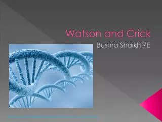
Watson and Crick model of DNA. Nucleotides are linked by phosphodiester bonds to form polynucleotides. Phosphodiester bond Covalent bond between the phosphate group (attached to 5’ carbon) of one nucleotide and the 3’ carbon of the sugar of another nucleotide.

E N D
Nucleotides are linked by phosphodiester bonds to form polynucleotides. Phosphodiester bond Covalent bond between the phosphate group (attached to 5’ carbon) of one nucleotide and the 3’ carbon of the sugar of another nucleotide. This bond is very strong, and for this reason DNA is remarkably stable. DNA can be boiled and even autoclaved without degrading! 5’ and 3’ The ends of the DNA or RNA chain are not the same. One end of the chain has a 5’ carbon and the other end has a 3’ carbon.
5’ end 3’ end
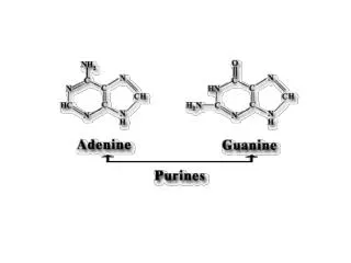
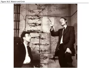
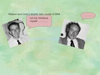
James D. Watson & Francis H. Crick - 1953 • Double Helix Model of DNA • Two sources of information: • Base composition studies of Erwin Chargaff • indicated double-stranded DNA consists of ~50% purines (A,G) and ~50% pyrimidines (T, C) • amount of A = amount of T and amount of G = amount of C (Chargraff’s rules) • %GC content varies from organism to organism Examples:%A %T %G %C %GC Homo sapiens 31.0 31.5 19.1 18.4 37.5 Zea mays 25.6 25.3 24.5 24.6 49.1 Drosophila 27.3 27.6 22.5 22.5 45.0 Aythya americana 25.8 25.8 24.2 24.2 48.4
James D. Watson & Francis H. Crick - 1953 Double Helix Model of DNA Two sources of information: X-ray diffraction studies - Rosalind Franklin & Maurice Wilkins Conclusion-DNA is a helical structure with distinctive regularities, 0.34 nm & 3.4 nm.
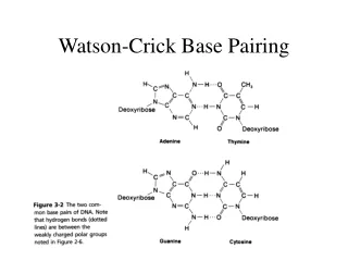
Double Helix Model of DNA: Six main features Two polynucleotide chains wound in a right-handed (clockwise) double-helix. Nucleotide chains are anti-parallel: 5’ 3’ 3’ 5’ Sugar-phosphate backbones are on the outside of the double helix, and the bases are oriented towards the central axis. Complementary base pairs from opposite strands are bound together by weak hydrogen bonds. A pairs with T (2 H-bonds), and G pairs with C (3 H-bonds). e.g., 5’-TATTCCGA-3’ 3’-ATAAGGCT-3’ Base pairs are 0.34 nm apart. One complete turn of the helix requires 3.4 nm (10 bases/turn). Sugar-phosphate backbones are not equally-spaced, resulting in major and minor grooves.
1962: Nobel Prize in Physiology and Medicine James D. Watson Francis H. Crick Maurice H. F. Wilkins What about? Rosalind Franklin