Download
1 / 40
400 likes | 566 Views
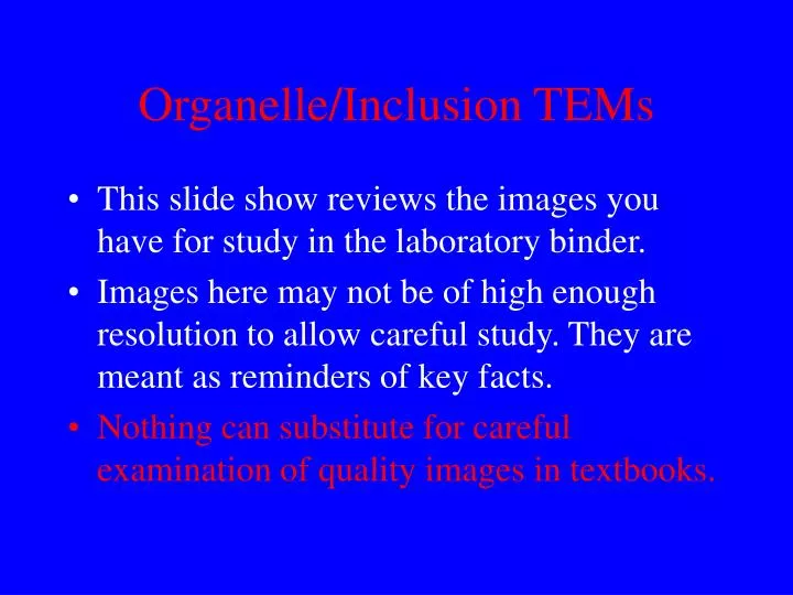
Organelle/Inclusion TEMs. This slide show reviews the images you have for study in the laboratory binder. Images here may not be of high enough resolution to allow careful study. They are meant as reminders of key facts.

E N D
Organelle/Inclusion TEMs • This slide show reviews the images you have for study in the laboratory binder. • Images here may not be of high enough resolution to allow careful study. They are meant as reminders of key facts. • Nothing can substitute for careful examination of quality images in textbooks.
Nucleus • This TEM depicts a typical nucleus. • Usually the largest cellular organelle. • Note both euchromatin and heterochromatin. • Can you ID other organelles here?
Nucleus • This cell appears to have multiple nuclei, but it does not. Why does the nucleus look like this? • Remember that while most cells have only 1 nucleus, some do indeed have more.
Nucleus • This close-up view shows euchromatin surrounding heterochromatin. • Do not mistake these blobs of nuclear material for glycogen particles.
Nucleus • Here, both the nucleolus and nuclear pores are evident. • Pores are indicated by the small arrows. • The surrounding RER and the large nucleolus indicate biosythesis of proteins.
Nucleus • Freeze-fracture reveals the distribution of pores in the nuclear envelope. • Pores are dynamic and can open and close. • We know little about how pores are positioned.
Mitochondria/Plasma Membrane • Here, mitochondria are localized to the basal cell cytoplasm. • They associate with the basal membrane in order to provide ATP for membrane located ATP-dependent ion pumps.
Mitochondria/Glycogen • The top panel shows a cell totally filled with mitochondria. • The lower panel shows mitochondria and numerous glycogen particles.
Mitochondria • These are typical mitochondria cut in cross-section. • Remember that this diameter is about 0.5 mm and use this as a reference value.
Mitochondria • Here, mitochondria are located between the filaments in cardiac muscle. • Contrast this location with mitochondria in skeletal muscle.
Mitochondria • This is a cross-section of cardiac muscle. • The mitochondria are seen as irregularly shaped objects. • Cristae are apparent and aid identification.
Smooth ER • This cytoplasm is also entirely filled with SER. • Some RER with ribosomes is present. • “L” indicates a lysosome. • Mitochondria are also seen.
Endoplasmic Reticulum • SER fills the space between mitochondria with tubular cristae in this cell. • What is this cell likely synthesizing? • “L” indicates lipid droplets.
Endoplasmic Reticulum • Smooth ER fills much of this cytoplasm. • “LF” indicates probable lipofuchsin material or a secondary lysosome. • Can you find a bit of the nucleus in this image?
Endoplasmic Reticulum • Rough ER studded with ribosomes. • Can you determine what areas are cytoplasmic and what areas are compartmentalized?
Endoplasmic Reticulum • RER showing attached ribosomes very clearly. • Note the polysomes sectioned transversely. These look like spirals, with one shown at the arrow.
Golgi Apparatus • A poorly organized Golgi complex is located beneath a centriole. • Many of the Golgi sacs are distended, probably because they were filled with protein for secretion.
Golgi/Endoplasmic Reticulum • A Golgi complex in the center of the field is surrounded by RER and SER. • The black dots are NOT glycogen, but packaged protein. (They are membrane enclosed.)
Golgi Apparatus • This Golgi complex shows very distended saccules. • Note the extensive RER surrounding it.
Glycogen Particles • Glycogen particles, or rosettes, are seen here along with SER and mitochondria. • Glycogen is a cellular inclusion, not a membrane limited organelle.
Lysosomes • Numerous secondary lysosomes are seen in the upper half of this figure. • Two peroxisomes are seen at the bottom. How are these identified?
Lysosomes/Peroxisomes • Both lysosomes and peroxisomes are shown in these panels. • Can you determine which is which? • Do you understand their respective functions?
Secretory Vescicles • Why are these identified as secretory vesicles (granules) and not lysosomes? • Can we be positive of our identification here? • Practice by looking at other images.
Organelle Review • Images such as this allow you to see many organelles at once. • Compare relative sizes, locations, number etc. • Is this cell making protein?
Organelle Review • Review images like this in you textbooks. • Find such images in chapters we have not yet covered and just concentrate on identifying organelles. • Training your eyes takes practice.
Organelle Review • Nuclei, plasma membranes and mitochondria should be apparent to you even at this low magnification. • Locate euchromatin and heterochromatin.
Organelle Review • Here, cells are very active in protein biosynthesis. • What criteria tell you this?
Organelle Review • This is a cell similar to those shown on the preceding image. • Note the distended saccules of RER and the euchromatic nucleus.
Microtubules • Seen best in the lower panel, MTs here radiate from a centriole. • They are straight, hollow tubes made of tubulin protomers assembled into protofilaments.
Centrioles • Centrioles are composed of MTs, in triplet spirals when seen in cross-section (left). • A longitudinal section of a centriole is to the right. • Function of centrioles?
Centrioles • Centrioles exist as pairs, oriented at right angles to each other. • Centrioles are NOT membrane-limited. • Basal bodies anchoring cilia are modified centrioles.
Microtubules • “A”= anaphase. MTs help separate the chromosomes. • “C” and “D” show different sections of the area indicated in “B”. MTs are cut in cross/longitudinal section (left/right).
MTs/Intermediate Filaments • Review this image at higher magnification in the laboratory. • MTs are hollow in cross-section and 25mm in diameter. • Ifs are not hollow and only 10 nm in diameter.
Actin Microfilaments • Microvilli contain a core of actin microfilaments • These filaments are anchored in the cytoplasm • Microvilli contain NO microtubules
Actin Microfilaments • Freeze-etch of actin microfilaments in microvilli. • Their interaction with the internal terminal web is easily seen using this method.
Mitosis • Make sure that you can identify cells in interphase, prophase, metaphase, anaphase and telophase.
Prophase • A cell in late prophase with the chromosomes not quite yet forming a true metaphase plate. • Could you have determined these were chromosomes and not, for example, mitochondria?
Metaphase • A good metaphase plate seen in transverse section using the TEM at low magnification. • You can get an impression of the MTs forming the spindles fibers here as well.
Telophase • A cell in telophase with a clear cleavage furrow (arrows). • Telophase is rapid and cells at this stage are difficult to find. • Anaphase is a much more lengthy process.
Conclusion • You should be able to identify all organelles and inclusions at the level of the electron microscope. • Some organelles and inclusions should be visible and identified using the LM. • Knowledge of molecular composition will also allow you to ID these structures. • Know the stages of mitosis.