Download
1 / 69
700 likes | 1.12k Views
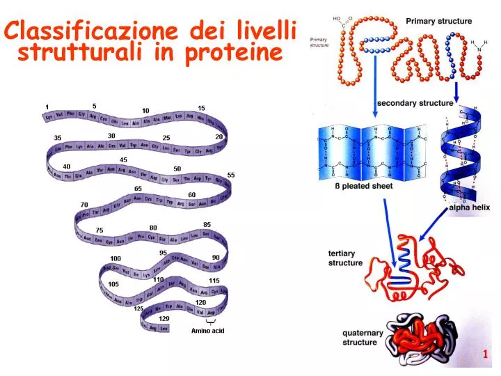
Classificazione dei livelli strutturali in proteine. Struttura primaria (sequenza amminoacidica ). Struttura primaria (sequenza amminoacidica ). struttura secondaria: la forma della catena principale della proteina ( protein backbone ). DIAGRAMMA DI RAMACHANDRAN.

E N D
Struttura primaria (sequenza amminoacidica)
Struttura primaria (sequenza amminoacidica)
struttura secondaria: la forma della catena principale della proteina (proteinbackbone)
DIAGRAMMA DI RAMACHANDRAN Gopalasamudram Narayana Iyer Ramachandran 8 October 1922 - 7 April 2001
DIAGRAMMA DI RAMACHANDRAN Gopalasamudram Narayana Iyer Ramachandran 8 October 1922 - 7 April 2001 Journal of Molecular Biology - 1963
DIAGRAMMA DI RAMACHANDRAN 180 φ 0 ψ ψ φ 0 180 -180
Conformazione delle catene laterali
eliche α in α-helix hydrogen bonds between carbonyl i and amide i+4 An α-helix in ultra-high-resolution electron density contours, with O atoms in red, N atoms in blue, and hydrogen bonds as green dotted lines (PDB file 2NRL, 17-32).
180 eliche α, ma non solo 0 ψ φ 0 180 -180
Strutture supersecondarie o motivi (elementi di struttura secondaria uguali o diversi si combinano) a: bab b: ripiegamento a forcina a: motivo aa d: barile b e: barile a/b (e)
Domini strutturali (subunità avente stabilità strutturale ed una propria funzione) dominio che lega il gliceraldeide-3-fosfato dominio che lega il NAD+ gliceraldeide-3-fosfato deidrogenasi
Enzima bifunzionale Piruvato chinasi PRA-isomerasi IGP-sintetasi
Struttura quaternaria Eteromultimeri Omomultimeri
Intrinsically Unstructured Proteins (IUPs) Set of keywords used: Natively denatured Natively unfolded Intrinsically unstructured Intrinsically disordered The Increase in the Number of PubMed Hits Dealing with Intrinsically Unstructured Proteins
Implications… Cell size constraints? The natively unstructured state is a simple and elegant solution adopted by evolution to avoid large protein, genome and cell sizes TiBS, 2003, 28, 81 Schematic representation of dimers (a) unstable (disordered) and (b) stable (ordered) monomers. Although in both cases the interface area between the monomers is the same, the size of the ordered monomer is much larger compared with the disordered example
Some Structural Features Combination of low overall hydrophobicity and relative high net charge under physiological conditions A combination of low mean hydrophobicity and high net charge preclude the formation of a hydrophobic cluster and promote an extended conformation Blue squares=275folded Red circle=91 IUPs Green circles=130 Predicted IUPs Cyan circle=242 Homologues of IUPs Proteins, 2000, 41,415
Some Structural Features… IUPs have a distinctive aa composition Table 2 Aminoacid Frequencies of Ordered and Disordered Proteins IUPs are enriched in S,P,E,K (disorder promoting) and depleted in W, Y,F,C,I, L, N (order promoting) TiBS, 2002, 527
This three-dimensional map of the protein universe shows the distribution in space of the 500 most common protein folds as represented by spheres. The spheres, which are colored according to classification, reveal four distinct classes. From http://www.lbl.gov/Science-Articles/Archive/PBD-Universe-map-Kim.html It is conceivable that, of the primordial peptides, those containing fragments with high helix and/or strand propensity found their way to fold into small alpha, beta, and alpha plus beta structures," Kim says. "The alpha slash beta fold structures do not appear until proteins of sufficient size rose through evolution and the formation of supersecondary structural units became possible. estimated number of protein folds: ~ 2000 (?)