Download
1 / 55
550 likes | 560 Views
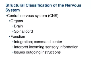
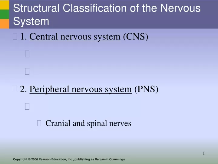
Structural Classification of the Nervous System. 1. Central nervous system (CNS) 2. Peripheral nervous system (PNS) Cranial and spinal nerves. Functions of the Nervous System. 1. Sensory input – (PNS) 2. Integration (CNS) 3. Motor output (PNS).

E N D
Structural Classification of the Nervous System • 1. Central nervous system (CNS) • 2. Peripheral nervous system (PNS) • Cranial and spinal nerves
Functions of the Nervous System 1. Sensory input – (PNS) 2. Integration (CNS) 3. Motor output (PNS)
Functions of Peripheral Nervous System (3 slides) • Sensory (afferent) division Figure 7.1
Functions of Peripheral Nervous System • Motor (efferent) division • Two subdivisions - Somatic nervous system - Autonomic nervous system- 2 subdivisions • Sympathetic • Parasympathetic Figure 7.1
Organization of the Nervous System Figure 7.2
Compare/Contrast Motor Divisions SOMATIC NS AUTONOMIC NS 2 branches – sympathetic and parasympathetic Uses acetylcholine, epinephrine, norepinephrine
Comparison of Somatic and Autonomic Nervous Systems Figure 7.24
Autonomic NS Branches • Parasympathetic- • Remember as the “D” division - digestion, defecation, and diuresis • Sympathetic – • Remember as the “E” division = extreme exercise, excitement, emergency, and embarrassment ** Both systems work in conjunction with one another to maintain homeostasis**
Types of Supporting Cells • Neuroglia – Nerve Glue (supporting cells) • AKA – glial cells • Types • Astrocytes • Microglia • Ependymal • Oligodendrocytes • Schwann cells and Satellite Cells (PNS)
CNS: Support Cells • Astrocytes • Microglia • Spider-like phagocytes Figure 7.3a
CNS: Support Cells • Ependymal cells • Oligodendrocytes • Producemyelin sheath around nerve fibers Figure 7.3b–c
PNS: Support Cells(2 major types) • 1. Satellite cells • 2. Schwann cells Figure 7.3e
Nervous Tissue: Neurons • Neurons = nerve cells • Major parts of a neuron • Cell body – • Processes –
Neuron Anatomy • Cell body • Nissl substance • Neurofibrils – Figure 7.4a
Neuron Anatomy • Processes - • Dendrites – • Axons – Figure 7.4a
Axons and Nerve Impulses Axons (cont’d) • Synaptic cleft • Synapse
Nerve Fiber Coverings • Schwann cells – ? • Nodes of Ranvier – • Oligodendrocytes - ? Figure 7.5
Vocab:CNS • CNS– contains mostly cell bodies • Gray matter – • Tracts - • White matter –
Functional Classification of Neurons (3) • 1. Sensory (afferent) neurons (PNS) • Special Sense • Cutaneous sense organs • Proprioceptors – • 2. Association neurons (interneurons) • CNS • 3. Motor (efferent) neurons
Neuron Classification Figure 7.6
Video : Review “Creation of a Nerve Impulse” • Video: Review “Nerve impulse propagation”
4 Main Regions of the Brain • 1. Cerebral hemispheres • 2. Diencephalon • 3. Brain stem • 4. Cerebellum • Brain song Figure 7.12b
Cerebral Hemispheres (Cerebrum) Cerebrum • Gyri (pl) – • Sulci (pl) – • Fissures – Figure 7.13a
Lobes of the Cerebrum • Lobes of the cerebrum • Frontal lobe • Parietal lobe • Occipital lobe • Temporal lobe • Insula Insula
Layers of the Cerebrum 3 slides • Cerebral cortex (Gray matter) Figure 7.13a
Specialized Areas of the Cerebrum Speech, memory, consciousness, emotional and logical response, voluntary movement, interpretation of sensation Figure 7.13c
Layers of the Cerebrum games • Cerebral White matter • Ex: corpus callosum connects hemispheres Figure 7.13a
Layers of the Cerebrum • Basal nuclei (basal ganglia) – internal islands of gray matter Figure 7.13a
Diencephalon – “interbrain” Diencephalon • Made of three parts • Thalamus • Hypothalamus • Epithalamus
Diencephalon: Thalamus Thalamus
Diencephalon: Hypothalamus Hypothalamus • Important autonomic n.s. center • part of the limbic system • Mammillary bodies (smell) hang off of hypothalamus
Diencephalon: Epithalamus Epithalamus • Houses the pineal body (an endocrine gland-sleep/wake cycles)
Review location/function on diencephalon Figure 7.15a
Brain Stem Parts of the brain stem • Midbrain • Pons • Medulla oblongata
Midbrain • Mostly tracts of nerve fibers • Cerebral peduncles- • ascend and descend impulses • Corpora quadrigemina -
Pons(“bridge”) • Below midbrain
Medulla Oblongata Contains vital visceral? control centers
Reticular formation – gray matter running length of brain stem • RAV – reticular activating system
Cerebellum • balance and equilibrium
Protection of the Central Nervous System Figure 7.16a
Meninges – 3 layers • Dura mater – “tough or hard mother” • Periosteum – • Meningeal layer – • Arachnoid layer • Arachnoid villi – • Pia mater (deepest)
Cerebrospinal Fluid (CSF) • Similar to blood plasma • Formed by the choroid plexus? (epithalamus) • Hydrocephalus – • Corrected by shunts
Blood Brain Barrier • Composed of the least permeable capillaries of the body Useless against some substances • Glucose and water • Fats and fat soluble molecules • Respiratory gases • Alcohol • Nicotine • Anesthesia