Download

1 / 49
490 likes | 619 Views
Chapter 38 Reproduction and Development (Sections 38.5 - 38.8). 38.5 Hormones and the Menstrual Cycle. Hormones coordinate cyclic changes in ovaries and uterus Cyclic changes in the uterus ( menstrual cycle ) begin with the onset of menstruation menstrual cycle

E N D
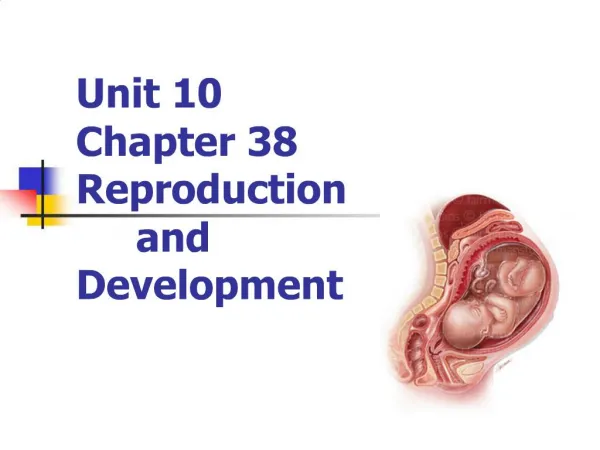
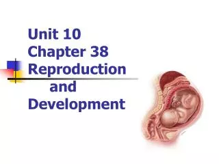
Chapter 38Reproduction and Development(Sections 38.5 - 38.8)
38.5 Hormones and the Menstrual Cycle • Hormones coordinate cyclic changes in ovaries and uterus • Cyclic changes in the uterus (menstrual cycle) begin with the onset of menstruation • menstrual cycle • Approximately 28-day cycle in which the uterus lining thickens and then, if pregnancy does not occur, is shed • menstruation • Flow of shed uterine tissue out of the vagina
Ovarian and Menstrual Cycles 1. Menstrual cycle begins: GnRH secretion by the hypothalamus causes the pituitary to increase output of follicle-stimulating hormone (FSH) and luteinizing hormone (LH) 2. FSH stimulates an ovarian follicle to begin maturing 3. As a follicle matures, cells around the oocyte secrete estrogens (the interval of follicle maturation preceding ovulation is the follicular phase of the menstrual cycle)
Ovarian and Menstrual Cycles (cont.) 4 . Estrogens bind to cells of the endometrium and signal to begin cell divisions (mitosis) that thicken the endometrium 5. The pituitary detects increased level of estrogens in the blood and responds with a surge of LH production 6. The midcycle surge of LH causes the primary oocyte to complete meiosis I and undergo cytoplasmic division, and causes the follicle to swell and burst (ovulation )
Ovarian and Menstrual Cycles (cont.) 7. Immediately after ovulation, estrogen level declines until the corpus luteum forms (luteal phase of the cycle); it secretes some estrogen and a lot of progesterone • Estrogen and progesterone keep the hypothalamus from secreting FSH, so no other follicles can start to mature 8. Estrogens and progesterone also cause the uterine lining to thicken and encourage blood vessels to grow through it • The uterus is now prepared for a new pregnancy
Ovarian and Menstrual Cycles (cont.) 9. If pregnancy does not occur, the corpus luteum persists for about twelve days, then breaks down – estrogen and progesterone levels drop • The hypothalamus senses this decline, and stimulates the pituitary to increase secretion of FSH and LH • In the uterus, decline in estrogens and progesterone causes endometrial lining to break down, and menstruation begins
Hormones, Ovary, and Uterus Fig. 38.9a, p. 636
Hormones, Ovary, and Uterus hypothalamus GnRH anterior pituitary FSH LH A FSH and LH levels in blood 5 1 FSH LH FSH and LH stimulate follicle maturation LH surge triggers ovulation Fig. 38.9a, p. 636
Hormones, Ovary, and Uterus Fig. 38.9b, p. 636
Hormones, Ovary, and Uterus B Follicle changes in an ovary corpus luteum forms 6 corpus luteum breaks down 2 follicle matures ovulation corpus luteum secretes estrogens, progesterone follicle secretes estrogens Fig. 38.9b, p. 636
Hormones, Ovary, and Uterus Fig. 38.9c, p. 636
Hormones, Ovary, and Uterus C Estrogen and progesterone levels in blood Progesterone 7 Estrogens 3 estrogens, progesterone, cause uterine lining to thicken 9 low estrogen Fig. 38.9c, p. 636
Hormones, Ovary, and Uterus Fig. 38.9d, p. 636
Hormones, Ovary, and Uterus 8 D Changes in uterine lining menstrual flow 4 0 2 4 6 8 10 12 14 16 18 20 22 24 26 28 Days of cycle Follicular phase Luteal phase Fig. 38.9d, p. 636
Animation: Correlation Between Hormonal Levels and Cyclic Ovarian and Uterine Changes
Menstrual Disorders • Premenstrual Syndrome (PMS) • Increased aldosterone secretion stimulates reabsorption of sodium (and water) causing tissues to swell • Cycle-induced changes also cause depression, irritability, anxiety, headaches, and can disrupt sleep • Menstrual Pain • Prostaglandins stimulates uterine contractions (cramps) • Misplaced endometrial tissue (endometriosis) and benign uterine tumors (fibroids) also causes painful menstruation
From Puberty to Menopause • At puberty, increased estrogen secretion results in development of female secondary sexual traits (breasts) • A woman enters menopausewhen all follicles in her ovaries have either been released during a menstrual cycle or disintegrated as a result of normal aging • menopause • Permanent cessation of menstrual cycles • Hormonal changes may cause hot flashes
38.6 When Egg and Sperm Meet • Egg and sperm may meet as a result of sexual intercourse • For males, intercourse requires an erection – aging, circulatory problems, and smoking affect erectile function • When a woman becomes sexually excited, glands in the cervix secret mucus, and glands on the labia produce a lubricating fluid
Sexual Intercourse • Intercourse increases sympathetic stimulation • Oxytocin acts on the amygdala to inhibit fear and anxiety • During orgasm, endorphins flood the brain; a surge of oxytocin causes rhythmic muscle contractions; and in males, ejaculation forces semen out of the penis
Fertilization • Ejaculation puts about 300 million sperm (which can live for about three days) into the vagina • Sperm swim upward into the oviduct, where fertilization of a secondary oocyte, released at ovulation, usually occurs • The secondary oocyte is covered with follicle cells over a layer of secreted proteins that form a jelly coat around it • The plasma membrane of the sperm’s head has receptors that bind species-specific proteins in the jelly
Fertilization (cont.) • Binding of sperm to egg proteins triggers the release of protein-digesting enzymes from the cap on the sperm’s head • Receptors in oocyte’s plasma membrane bind a sperm’s plasma membrane, the two membranes fuse, and the sperm enters the secondary oocyte • The egg’s jelly coat changes to prevent other sperm from binding
Fertilization (cont.) • Binding of sperm and oocyte membranes initiates meiosis II in the secondary oocyte – unequal cytoplasmic division produces a single mature egg (ovum) and a polar body • Chromosomes in the haploid egg and sperm nuclei become the genetic material of the new zygote • ovum • Mature animal egg
Fertilization Occurs in the Oviduct Fertilization oviduct ovary Ovulation uterus opening of cervix A Fertilization most often occurs in the oviduct. Many human sperm travel swiftly through the vaginal canal into oviducts (blue arrows). Inside an oviduct, the sperm surround a secondary oocyte that was released from an ovary at ovulation. vagina sperm enter vagina Fig. 38.10a, p. 639
Enzymes Clear a Path for Sperm follicle cell jelly layer oocyte nucleus completing meiosis II polar body B Enzymes released from the cap of each sperm clear a path through the jelly layer. Penetration of the secondary oocyte by a sperm causes the oocyte to releases substances that harden the jelly layer and prevent other sperm from binding. Fig. 38.10b, p. 639
polar bodies haploid egg and sperm nuclei jelly layer C The oocyte nucleus completes meiosis II, and the oocyte undergoes unequal cytoplasmic division yielding a mature haploid ovum and a second polar body. The sperm’s tail and organelles degenerate, leaving only a haploid nucleus with the paternal genome. The nuclear membranes of the egg and sperm nuclei will break down and the paternal and maternal chromosomes will become arranged on a bipolar spindle in preparation for the first mitotic division of the new zygote. The photo at right is a light micrograph taken one day after fertilization. Fig. 38.10c, p. 639
ANIMATION: Fertilization To play movie you must be in Slide Show Mode PC Users: Please wait for content to load, then click to play Mac Users: CLICK HERE
ANIMATION: Menstrual cycle summary To play movie you must be in Slide Show Mode PC Users: Please wait for content to load, then click to play Mac Users: CLICK HERE
ANIMATION: Cell-mediated response To play movie you must be in Slide Show Mode PC Users: Please wait for content to load, then click to play Mac Users: CLICK HERE
ANIMATION: Hormones and the menstrual cycle To play movie you must be in Slide Show Mode PC Users: Please wait for content to load, then click to play Mac Users: CLICK HERE
ANIMATION: Implantation and Pregnancy To play movie you must be in Slide Show Mode PC Users: Please wait for content to load, then click to play Mac Users: CLICK HERE
38.7 Preventing Pregnancy • Couples can prevent pregnancy by avoiding sex entirely or abstaining when the woman is fertile • Hormones delivered by pills, patches, or injections can prevent a woman from ovulating • Temporary barriers such as condoms or a diaphragm keep sperm and egg apart, as do permanent surgical methods such as a vasectomy or tubal ligation
38.8 Sexually Transmitted Diseases • Each year, pathogens that cause sexually transmitted diseases (STDs) infect about 15 million Americans • Women are more easily infected than men, have more complications, can pass an STD on to her newborn • Both men and women can be debilitated by bodywide effects of some infections
Bacterial Diseases • The most common bacterial STD is now chlamydia (Chlamydia trachomatis), which often causes no symptoms in women, but can infect her child • Bacteria also cause gonorrhea (infect oviducts, causing cramps, scarring, and sterility in women), and syphilis (damages liver, bones, and eventually the brain) • Bacterial STDs can be cured with antibiotics
Complications of Pregnancy • Tubal pregnancy • Infant with chlamydia-inflamed eyes
Syphilis • Skin sores caused by untreated syphilis • Syphilis is caused by Treponema pallidum, a spiral-shaped bacteria
Human Papillomaviruses (HPV) • HPV infection is widespread in the United States • Of about 100 HPV strains, a few cause genital warts • Some strains of HPV cause cervical cancer • Sexually active females should have an annual Pap smear to screen cells for signs of cancer • Viral diseases cannot be cured by any drug • A vaccine (Gardasil) can prevent some types of HPV infection if given before viral exposure
Human Immunodeficiency Virus (HIV) • Infection by HIV can cause AIDS • Unprotected anal sex is 5 times more dangerous than unprotected vaginal sex and 50 times more dangerous than oral sex • If you may have been exposed to HIV, get tested as soon as possible – early treatment may prevent development of AIDS
Key Concepts • Human Sexual Behavior • Sexual intercourse brings together sperm and eggs, allowing fertilization • A variety of methods allow people who engage in intercourse to prevent fertilization. • Intercourse also raises the risk of infection by sexually transmitted bacteria, viruses, or protozoans