Download
1 / 98
980 likes | 1.06k Views
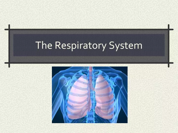
The Respiratory System. LUNG CAKE. Functions of the Respiratory System. Obtain O 2 from environment Expel CO 2 into the environment Filter foreign particle out of incoming air Regulates H 2 O content and temperature of incoming air Creates vocal sounds Contributes to sense of smell

E N D
Functions of the Respiratory System • Obtain O2from environment • Expel CO2 into the environment • Filter foreign particle out of incoming air • Regulates H2O content and temperature of incoming air • Creates vocal sounds • Contributes to sense of smell • Helps regulate blood pH
Steps for Respiration • 1) Breathing or ventilation • Moving air in and out of lungs • 2) External respiration • Exchange of gases between lungs and blood vessels • 3) Transport of gases to cells • 4) Internal respiration • Exchange of gases between blood vessels and cells • 5) Cellular respiration • Using O2 in metabolic processes to create energy
Divisions of Respiratory Tract • Upper respiratory tract • Nose • Nasal cavity • Paranasal sinuses • Pharynx • Lower respiratory tract • Larynx • Trachea • Bronchial tree • Lungs
Nose • Bone and cartilage • Support structure • Nostrils • Openings for entry of air • Nose hairs • Guard entrance and protect against particles
Nasal Cavity • Hollow space behind nose • Nasal septum • Bone and cartilage structure dividing cavity into 2 sides • Nasal conchae • Projections from nasal cavity wall; increase surface area • Lining of nasal cavity • Pseudostratifiedepithelial cells with mucous-secreting goblet cells • Particles are trapped and moved out and air is warmed and moistened
The NOSE bones and cartilage support nose, two openings (nostrils), hair filters large particles Nasal Cavity – hollow space behind the nose Nasal septum – divides the nose (bone)
Nasal conchae – bones that divide the nasal cavity, support the mucus membrane and increase surface area (superior, middle, inferior) * deviated septum – when the septum bends to one side
Function of the conchae - increase surface area Mucus Membrane - warms and moistens air, also traps particles (dust) *particles go to stomach Nasal Conchae
Paranasal Sinuses • Air-filled sacs within skull bones • Reduce the weight of the skull • Affect sound of the voice
Paranasal Sinuses - – spaces within the bones • maxillary • frontal • ethmoid • sphenoid reduce the weight of skull and are resonant chambers for voice.
Pharynx • Shared with digestive system • Divisions: • Nasopharynx • Oropharynx • Larygopharynx • Otherwise known as hypopharynx • Openings: • Oral cavity • Nasal cavity • Esophagus • Trachea
The three pharyngeal regions Pharynx – behind the oral cavity, between the nasal cavity and larynx (space, not a structure)
Larynx • Enlargement at beginning of trachea • Framework of muscle and cartilage bound by elastic tissue • Largest cartilages: • Thyroid (Adam’s apple) • Cricoid • Epiglottic
Vocal Cords • Housed in larynx • False vocal cords • Fold of tissue that helps to close passageway when swallowing • True vocal cords • Vibrate when air is forced through causing sound • Tension of chords helps regulate pitch • Force of air helps regulate volume of sound
Larynx – enlargement at the top of the trachea and below pharynx, conducts air in and out of trachea, houses vocal cords - composed of a framework of muscles and cartilages (thyroid (Adam’s apple), cricoids, epiglottic cartilages)
Glottis • Triangular opening between vocal cords • When swallowing: • False vocal cords close glottis • Larynx raises and presses epiglottis against opening
Glottis - false vocal folds (do not produce sound) – help close airway during swallowing - true vocal folds (produce sound) – changing shape of the pharynx, and oral cavity changes sounds into words - contracting and relaxing muscles changes pitch (increased tension = higher pitch)
www.voiceinfo.org Steven Tyler's Vocal Cords
Glottis – triangular slit that opens during breathing/talking, and closes during swallowing Epiglottis – flaplike structure that stands upright, allows air to enter larynx, during swallowing it presses downward and prevents food from entering air passages
LARYNGITIS When the mucus membrane becomes swollen and prevents the vocal cords from vibrating freely. Trachea (windpipe), flexible cylinder with cartilage to give it stiffness and keep it from collapsing Trachea leads to the BRONCHIAL TREE
Trachea • Also called windpipe • Descends anterior to esophagus • Lining of trachea: • Pseudostratifiedepithelial cells with mucous-secreting goblet cells • Enclosed with C-shaped rings of hyaline cartilage to prevent tube from collapsing
Bronchial Tree • Branching tubes connecting trachea to site of gas exchange in lungs • Right and left primary bronchi Secondary bronchi Tertiary bronchi Bronchioles Terminal bronchioles Respiratory bronchioles Alveolar ducts Alveolar sacs Alveoli • Amount of cartilage decreases through tract • Amount of smooth muscle in the walls increases through tract
Primary bronchii --> bronchioles --> alveolar ducts --> sacs --> alveoli *gas exchange
Alveoli • Terminal sacs at the end of the tract • Surrounded by network of blood capillaries • Site of gas exchange • Respiratory membrane: • Simple squamous epithelium in alveoli and blood capillary • Basement membrane fuses the two (alveoli and capillary) together
$20,000 PYRAMID! • A word/phrase will be posted – person A (who is facing the board) must attempt to explain the word (without saying it!) while Person B (who is NOT facing board) will guess what the word is. • NO DRAWING! • If your partner gets them all right – with NO CHEATING – raise your hands! • GET READY!!!! • Alveoli • Larynx • Trachae
Quick Quiz 1. What do you call the bones found within the nasal cavity? 2. What specific bone divides the nasal cavity into two sides? 3. The space at the back of the mouth is the________. 4. The spaces within the bones of the skull are called the ______________________ 5. What structure is known as the windpipe? ______ 6. What is the triangular slit that opens during breathing and talking? 7. In what structures does gas exchange occur? 8. During swallowing, this flap closes to prevent food from entering the airway: ______________________
Lungs • Located in thoracic cavity: • Mediastinum between • Thoracic cage surrounds • Diaphragm below • Suspended by bronchi and blood vessels • Cover by visceral pleura which folds back to become parietal pleura • Potential space between is pleural cavity • Small layer of serous fluid between • Right lung: 3 lobes • Left lung: 2 lobes
Right Lung = 3 lobes Left Lung = 2 lobes Serous fluid lubricates lungs during breathing
EXHALATION As the diaphragm and other muscles relax, ELASTIC RECOIL from surface tension forces air out. Muscles can force extra air out or in
Inspiration • At rest, pressure of air and thoracic cavity are the same; • NO net movement of air • Pressure must be decreased in thoracic cavity for air to move in • Diaphragm is contracted (lowered) and external (inspiratory) intercostal muscles contract as well • Contractions increase cavity volume which decreases pressure
Inspiration (cont) • Muscle movements pull on the pleural membranes to expand the lung • Surface tension • Difficulty in expanding alveoli because the walls tend to stick together • Surfactanthelps combat surface tension • Deeper breaths can be taken by contracting muscle more forcefully or by involving other muscles
Expiration • Normally passive process • Contracting muscles relax • Elastic recoil and surface tension cause tissue to return to previous state before inspiration • Pressure increases in cavity and forces air out of lungs