Download
1 / 10

E N D
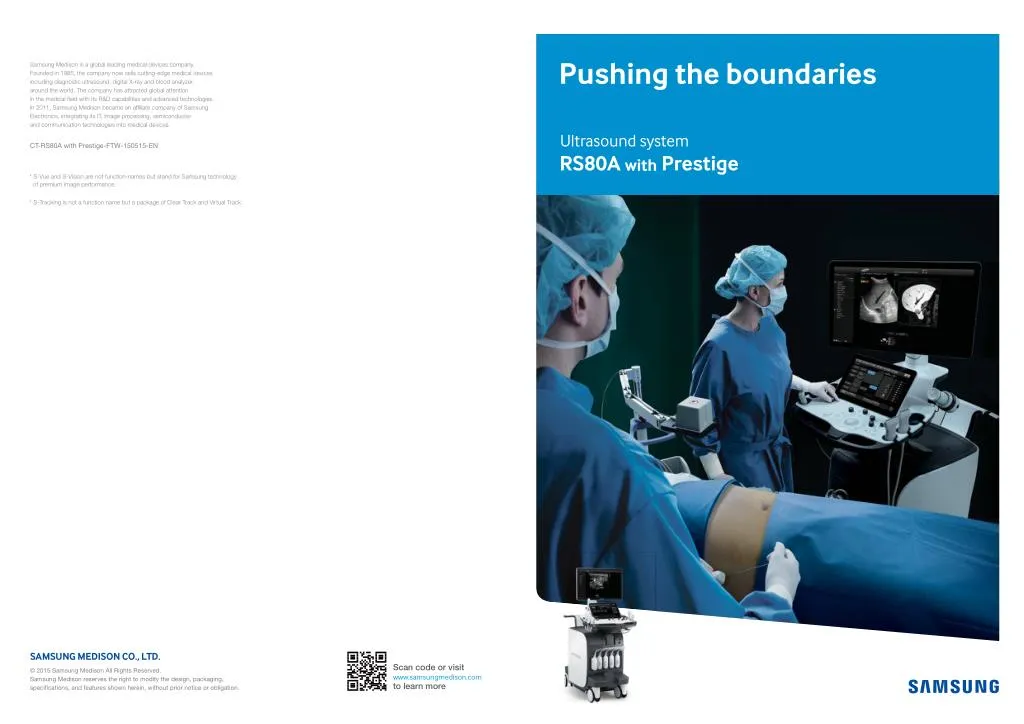
Pushing the boundaries Samsung Medison is a global leading medical devices company. Founded in 1985, the company now sells cutting-edge medical devices including diagnostic ultrasound, digital X-ray and blood analyzer, around the world. The company has attracted global attention in the medical field with its R&D capabilities and advanced technologies. In 2011, Samsung Medison became an affiliate company of Samsung Electronics, integrating its IT, image processing, semiconductor and communication technologies into medical devices. Ultrasound system CT-RS80A with Prestige-FTW-150515-EN RS80A with Prestige * S-Vue and S-Vision are not function-names but stand for Samsung technology of premium image performance. * S-Tracking is not a function name but a package of Clear Track and Virtual Track. SAMSUNG MEDISON CO., LTD. Scan code or visit www.samsungmedison.com to learn more © 2015 Samsung Medison All Rights Reserved. Samsung Medison reserves the right to modify the design, packaging, specifications, and features shown herein, without prior notice or obligation. Pushing the boundaries Samsung Ultrasound RS80A with Prestige I 2
????????????????????????????????????????????? Enhanced technologies expand capabilities Apr 2015 S-Vision beamformer The S-Vision beamformer demonstrates a clearer image that receives returning signals through a sophisticated digital filtering system resulting in reduced side lobes, less noise and artifact. It enhances the image quality with better clarity and consistent results. The advanced technical capabilities that the RS80A with Prestige features are built on the successes of Samsung technologies, including superior image quality, while offering exclusive options. The features such as S-Fusion, and S-Shearwave provide diagnostic confidence and user convenience in challenging practices. Conventional RS80A with Prestige S-Vision imaging engine Resolution Penetration Uniformity With the S-Vision imaging engine built on the RS80A with Prestige, the digital signals demonstrate clear, detailed resolution and tissue uniformity for various types of applications in general imaging. Noise S-Vue transducer (CA1-7A, CV1-8A) The S-Vue transducer provides a larger bandwidth and higher sensitivity both in transmit and receive capabilities. The combination of the new S-Vision Beamformer with the S-Vue transducer allows easier visualization of difficult-to- image pathologies. In addition, the ergonomically designed S-Vue transducer fits well in the hand and is easy to handle. Sensitivity Sensitivity S-Vue transducer Transmit Receive Transmit Receive f? f? 2f? 2f? Frequency Frequency Transmit/Receive at conventional transducer Transmit/Receive at S-Vue transducer * Compared with the conventional Samsung transducers Pushing the boundaries Samsung Ultrasound RS80A with Prestige I 4
Essential tools for interventional procedures Samsung pushes the boundaries of ultrasound technology. With leading technologies like S-Fusion and S-Tracking, you can expect accuracy in interventional procedures. S-Fusion S-Fusion enables simultaneous localization of a lesion with a real-time ultrasound image supported by other modalities' 3D-Datasets. Since the image fusion method still faces challenges such as relatively long registration time and low accuracy of registration, fusion speed and accuracy are the strength of Samsung's proprietary S-Fusion and it enables the system to be ready for advanced clinical applications. Apr 2015 Apr 2015 Apr 2015 Positioning Auto-registration S-Fusion with CEUS+ Registration time (Sec) S-Fusion imaging takes up to 66 seconds. Especially for Positioning Auto-registration, fusion imaging only takes about 30 seconds. It provides easy and fast registration by placing the transducer on the epigastrium and precise alignment, which allows you to focus on the interventional procedure. ??? 66 sec ?? 30 sec ? * Above result is an average value of internal tests. Manual Positioning Auto Apr 2015 S-Tracking S-Tracking increases the rate of accuracy during interventional procedures by providing the simulated path of the needle and the target mark in the live ultrasound image. Clear Track, one of two functions provided by S-Tracking, secures the accuracy by using a specialized needle with a sensor tip. Virtual Track uses general needles during the procedure, providing both accuracy and economic benefit. Clear Track * Above features may not available for use in some countries. Pushing the boundaries Samsung Ultrasound RS80A with Prestige I 6
Cutting-edge technology for diagnostic challenges With advanced technologies like S-Shearwave and CEUS+ thenumber of biopsies can be reduced, lesions become visible and examinations are easier to perform. S-Shearwave S-Shearwave detects the velocity of the shearwave propagated through the targeted lesion and displays the numerical measurement of stiffness in kPa or m/s together with a Reliable Measurement Index (RMI)*. Also it provides Variation Range (VR), a range of value, that intuitively shows the uniformity of tissue stiffness in the Region of Interest (ROI). The wider range means the less tissue stiffness uniformity. In the profile window, the user can easily edit each measurement value depending on its Reliable Measurement Index. S-Shearewave helps to reduce the number of conventional liver biopsies by providing quantitative tissue characteristic information. Shearwave Apr 2015 Apr 2015 Apr 2015 Variation Range ?????????????????????????????????????????????????????????????????????????????????????????????????????????????????????? the selection of optimal measurements. CEUS+ CEUS+ technology uses the unique properties of ultrasound contrast agents. When excited with a Low MI the oscillating micro bubbles reflect both the basic frequencies and harmonic signals. In the CEUS+ harmonic image on the left the perfused parts are displayed and on the right side a conventional B-Mode image. Apr 2015 Apr 2015 Liver metastasis needle biopsy Liver metastasis arterial phase * Above features may not available for use in some countries. Pushing the boundaries Samsung Ultrasound RS80A with Prestige I 8
Advances that minimize risk Advanced QuickScan™ Apr 2015 Advanced QuickScanTM technology provides intuitive optimization of gray scale and Doppler parameters. One touch of the QuickScanTM button elevates efficiency and workflow by adjusting functions including color gain and color box location. Early detection of cardiovascular diseases and risk for stroke Apr 2015 Apr 2015 CCA Doppler without QuickScan™ CCA Doppler with QuickScan™ Advanced QuickScanTM Apr 2015 Auto IMT+™ Auto IMT+™ is a screening tool that analyzes a patient's risk of stroke and heart disease. It allows easy intra-media thickness measurement of both the anterior and posterior wall of the common carotid by clicking a button. This simple procedure enhances exam productivity and adds diagnostic value. IMT (Intra-Media Thickness) measured with Auto IMT+™ Sample Volume Setting Angle Rotating ROI Positioning Arterial Analysis Strain+ Stress Echo Strain+ quantitatively displays a Bull's Eye which shows left ventricular motion and dyssynchrony at a glance. The Stress Echo package includes wall motion scoring and reporting. It includes exercise Stress Echo, pharmacologic Stress Echo, diastolic Stress Echo and free programmable Stress Echo. Arterial Analysis detects functional changes of vessels, providing measurement values such as the stiffness and intra-media thickness. Since the functional changes occur before morphological changes, this technology supports the early detection of cardiovascular diseases. Apr 2015 Apr 2015 Apr 2015 Augmentation index Measurement table of Arterial Analysis Strain+ Stress Echo * Above features may not available for use in some countries. Pushing the boundaries Samsung Ultrasound RS80A with Prestige I 10
??????????????????????????????????????? Apr 2015 For a better ultrasound breast assessment??????????????? awide range of useful imaging and quantitative tools E-Breast™ (ElastoScan™ for breast) S-Detect™ for breast S-Detect™ employs Breast Imaging-Reporting and Data System (BI-RADS®) scores for standardized analysis and classification of suspicious lesions. By simply clicking the suspected area, it draws the lesion area and provides the characteristics of the lesion and a recommendation on whether the lesion is benign or malignant. Such technology assists in a more accurate diagnosis, while improving the efficiency of workflow and reducing the time users spend in repetitive tasks. Apr 2015 E-Breast™ is a technology that calculates the strain ratio between the selected target and surrounding fatty tissues. Unlike conventional ultrasound elastography, E-Breast™ requires only one ROI to be selected by the user. This simplified process enhances consistency and reduces the chance of error by eliminating the step of manual selection of the surrounding fatty tissue region. Apr 2015 Breast Parenchyma Volume linear transducer Apr 2015 Multi-dimensional volume data acquired by Volume linear transducer visualizes the structure of targeted planes in a single step. It helps users to deliver more accurate and efficient diagnosis. Exam report with S-DetectTM Fybro adenoma * Above features may not available for use in some countries. Pushing the boundaries Samsung Ultrasound RS80A with Prestige I 12
Superior image clarity with enhanced technology Apr 2015 Apr 2015 Apr 2015 Apr 2015 Kidney transplantation GB stones Thyroid nodule Thyroid nodule Apr 2015 Apr 2015 Apr 2015 Apr 2015 Wrist ganglion Pediatric spine Finger ganglion Quadriceps Apr 2015 Apr 2015 Apr 2015 Apr 2015 Carotid artery Panoramic 4 Chambers Wrist vessel Pushing the boundaries Pushing the boundaries Samsung Ultrasound RS80A with Prestige I 14 Samsung Ultrasound RS80A with Prestige I 14
Uncompromising quality and ease of use Count on Samsung for an easy alliance between user and technology. Streamlined processes with EZ-Exam+TMand the convenience provided by Natural Vue ensure an optimum user experience. Apr 2015 E-Thyroid™ (ElastoScan™ for thyroid) Apr 2015 E-Thyroid™ provides an assessment of thyroid lesions by incorporating an index for suspicious areas. E-Thyroid™ images are generated using pulsations from the adjacent Carotid Artery, eliminating the need for manual transducer compression and offering greater consistency. Thyroid ElastoScanTM nodule Apr 2015 Natural Vue The 3D Natural Vue delivers a realistic view of the surface. It offers morphological information, including form, size and location of the Region of Interest (ROI) compared to 2D images. GB mass in Natural Vue * Image provided by JY Lee at SNUH Apr 2015 EZ-Exam+TM EZ-Exam+™ transforms multiple ultrasound investigation steps into a streamlined process. It enables users to build a fast and convenient diagnostic environment by storing optimized, preferred protocols with the EZ-Exam+™ function control. Set up display of EZ-Exam+™ * Above features may not available for use in some countries. Pushing the boundaries Samsung Ultrasound RS80A with Prestige I 16
Designed for your convenience Folding monitor 23-inch LED display The RS80A with Prestige features a 23-inch high definition LED display delivering excellent contrast resolution, image clarity and vibrant color in any lighting condition. The folding monitor enables safe and secure transport. Apr 2015 Apr 2015 13.3-inch tilting touch screen Simplified console design The tilting touch screen adjusts to accommodate user viewing preference in any scanning environment. The simplified control panel including 3D Navigator and intuitive grouping of console buttons streamlines system interaction for efficient patient scanning. 6 way adjustable control panel Swivel lock The RS80A with Prestige's 6 way adjustable control panel optimizes the work environment to reduce repetitive stress. Upon power down the control panel returns to home position for easier mobility. A single pedal controls a swivel lock mechanism to conveniently secure the console in place and accommodates efficient movement during a variety of scanning procedures. Pushing the boundaries Samsung Ultrasound RS80A with Prestige I 18
Comprehensive selection of transducers Curved array transducers Volume transducers * S-Vue transducer * S-Vue transducer CA1-7A CA2-8A CF4-9 CV1-8A V5-9 V4-8 LV3-14A • Application : pediatric, vascular • Application : abdomen, obstetrics, gynecology, contrast • Application : abdomen, obstetrics, gynecology • Application : abdomen, obstetrics, gynecology • Application : obstetrics, gynecology, urology • Application : abdomen, obstetrics, gynecology • Application : musculoskeletal, small parts, vascular • Field of View : 92° • Field of View : 58° • Field of View : 72° • Field of View : 76° • Field of View : 38.4mm • Field of View : 70° • Field of View : 150.6° Linear array transducers Endocavity transducer Phased array transducers L3-12A LA3-16A LA2-9A E3-12A PM1-6A PA3-8B PA4-12B LA4-18B • Application : small parts, vascular, musculoskeletal • Application : small parts, vascular, musculoskeletal • Application : small parts, vascular, musculoskeletal • Application : small parts, vascular, musculoskeletal, abdomen • Application : obstetrics, gynecology, urology • Application : cardiac, TCD, abdomen • Application : cardiac, pediatric, abdomen • Application : cardiac, pediatric • Field of View : 90° • Field of View : 210° • Field of View : 37.5mm • Field of View : 50mm • Field of View : 38.4mm • Field of View : 44.16mm • Field of View : 90° • Field of View : 90° CW transducers CW6.0 DP2B L7-16 LA3-16AI • Application : small parts, vascular, musculoskeletal • Application : musculoskeletal • Application : cardiac • Application : cardiac • Field of View : 25.6mm • Field of View : 38.4mm * Above options may not available for use in some countries. Pushing the boundaries Samsung Ultrasound RS80A with Prestige I 20