Download
1 / 38
1.37k likes | 3.52k Views
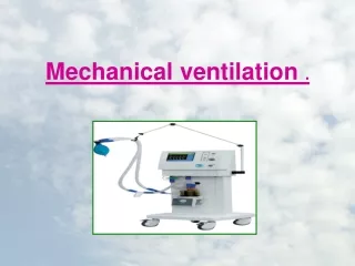
Basics of Mechanical Ventilation. Origins of mechanical ventilation. Negative-pressure ventilators (“iron lungs”) first used in Boston Children’s Hospital in 1928 Used extensively during polio outbreaks in 1940s – 1950s.

E N D
Origins of mechanical ventilation • Negative-pressure ventilators (“iron lungs”) • first used in Boston Children’s Hospital in 1928 • Used extensively during polio outbreaks in 1940s – 1950s The iron lung created negative pressure in abdomen as well as the chest, decreasing cardiac output. Iron lung polio ward at Rancho Los Amigos Hospital in 1953.
Era of intensive care begun with this • Positive-pressure ventilators • Invasive ventilation first used at Massachusetts General Hospital in 1955 • Now the modern standard of mechanical ventilation
Outline • Modes • Ventilator Settings • Indications to intubate • Indications to extubate • Trouble shooting
Pressure ventilation vs. volume ventilation Pressure-cycled modes: -deliver a fixed pressure at variable volume Volume-cycled modes: -deliver a fixed volume at variable pressure
Ventilator settings • Ventilator mode • Respiratory rate • Tidal volume or pressure settings • Inspiratory flow • I:E ratio • PEEP • FiO2 • Inspiratory trigger
CMV-Volume Tidal Volume Volume
Pressure Support Ventilation (PSV) Patient determines RR, VE, inspiratory time – a purely spontaneous mode
CPAP and BiPAP CPAP is essentially constant PEEP; BiPAP is CPAP plus PS • Parameters • CPAP – PEEP set at 5-10 cm H2O • BiPAP – CPAP with Pressure Support (5-20 cm H2O) • Shown to reduce need for intubation and mortality
Respiratory Rate • 10-12/Min – Adult • 20+_ 3 - Child • 30- 40 - New born
Respiratory Rate • Increase – Hypoxia Hypercapnoea / Resp.Acidosis • Decrease Hypocapnoea Resp.Alkalosis Asthma / COPD
Tidal Volume or Pressure setting • Optimum volume/pressure to achieve good ventilation and oxygenation without producing alveolar overdistention • Max = 6-8 cc/kg
Inspiratory Trigger • Normally set automatically • 2 modes: • Airway pressure • Flow triggering
I:E Ratio • Normaly 1:2 • Asthma/COPD 1:3, 1:4, … • Severe hypoxia ARDS/ALI Pul.Edema 1:1 , 2:1
FIO2 • Goal – to achive PaO2 > 60mmHg or a sat >90% • Start at 100% aim 40%
Vent settings to improve <oxygenation> PEEP and FiO2 are adjusted in tandem • FIO2 • Simplest maneuver to quickly increase PaO2 • Long-term toxicity at >60% • Free radical damage • Inadequate oxygenation despite 100% FiO2 usually due to pulmonary shunting • Collapse – Atelectasis • Pus-filled alveoli – Pneumonia • Water/Protein – ARDS • Water – CHF • Blood - Hemorrhage
Positive End-expiratory Pressure (PEEP) What is PEEP? Positive pressure measured at the end of expiration. What is the goal of PEEP? • Improve oxygenation • Recruit lung in ARDS • Prevent collapse of alveoli • Diminish the work of breathing
PEEP- Indications. • If a PaO2 of 60 mmHg cannot be achieved with a FiO2 of 60% • If the initial shunt estimation is greater than 25% • Pulmonary edema • ARDS/ALI • Atelectosis
PEEP • What are the secondary effec`ts of PEEP? • Barotrauma • Diminish cardiac output • Regional hypoperfusion • Augmentation of I.C.P.? • Paradoxal hypoxemia • Hypercapnoea and respiratory acidosis
PEEP • Contraindication: • Barotrauma • Airway trauma • Hemodynamic instability • I.C.P.? • Bronchospasm?
Collapse/ atelectosis/ ARDSIncreases Surface area for gas exchangeOpens the collapsed lung PEEP Collapsed alveoli After PEEP
Pulmonary edemaTranslocation of fluid to peribroncheal region – helps in oxygenation PEEP
DOPE • D- Disposition of ETT • O- Obstruction / kinking • P- Pneumothorax • E- Equipment failure
Need for tracheostomy Prolonged intubation may injure airway and cause airway edema 1 - Vocal cords. 2 - Thyroid cartilage. 3 - Cricoid cartilage. 4 - Tracheal cartilage. 5 - Balloon cuff.