Download
1 / 21
220 likes | 342 Views
Anatomy and Physiology For The First Class 2 nd Semester. The Special Senses. The Special Senses. Five special Senses Olfection (smell) Gustation (taste) Vision (sight) Hearing Equilibrium. Vision . Vision is extremely important to human survival.

E N D
Anatomy and Physiology For The First Class 2nd Semester
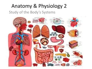
The Special Senses • Five special Senses • Olfection (smell) • Gustation (taste) • Vision (sight) • Hearing • Equilibrium
Vision Vision is extremely important to human survival. More than half the sensory receptors in the human body are located in the eye. Structurally two eyes are separate but some of their activities are coordinated so that they function as a pair. It is possible to see with only one eye, but three dimensional vision is impaired especially in the judgment of speed and distance. Accessory structures of the eye include the eyelids, eyelashes, eyebrows, lacrimal (tearing) apparatus, and extrinsic eye muscles.
Lids and lashes: help to protect eyes. • Blink reflex: closes the eyelids when environmental hazards reach near the eye. • Tears are necessary to keep eyes moist and healthy. They produce smooth surface (important for clear vision), provide Oxygen and other nutrients to the surface of the eye and contain proteins like lysozymes, immunoglobulin, lactoffrin which all helps to ward off infection. • Lacrimal gland and small other glands inside the eyelid constantly produce tears to keep eye moist. • Tears drain from the eye through two small opening called upper and lower puncta located inside upper and lower corner of eyelid near the nose.
The most visible structures of the eyes • Sclera: is a white part of the eye, composed of thought fibrous layer. sclera helps to protect and support eyeball. • Cornea: is an anterior extension of sclera, it is very transparent, therefore it is invisible. • Pupil: is the dark central window into the anterior of the eye. Image we see result from the light enters through the pupil. • Iris: is the colored part around the pupil. Muscles of the iris change the size of the pupil opening making the pupil larger in the dim light and smaller in the bright light. • Conjunctiva: is the transparent layer of tissue ( mucous membrane) covers the eye (except the cornea) and inside the eyelids. The conjunctiva produces mucus that helps to protect and lubricate the eye and lids.
Muscles of the eye • Superior rectus muscle • Inferior rectus muscle • Medial rectus muscle • Lateral rectus muscle • Superior oblique muscle • Inferior oblique muscle
Cross Section of the Eye (Structure of Eyeball) • The greater part of eyeball (posterior 5/6) is a sphere in shape. • The anterior 1/6 is much more convex than the posterior part. • The wall of eyeball is made up of three main layers: • The outer layer is called the Fibrous layer: the anterior part is transparent and is called Corneawhile the reminder is called Sclera. • The second layer is theVascular layer (Uvea): consists of: 1). Choroid2). Ciliary body 3).Iris. • The innermost layer is called the Retina. It contains photoreceptors that convert the stimulus of light into nervous impulses, these receptors are of two kinds, Rods and Cons. there are about 7 million cons in each retina. The rods are more numerous 100 million in number. • There are three chambers the anterior chamber which is located between the cornea and the iris (contains aqueous humor), the posterior chamber located between the iris and the front of the lens and the vitreous chamber located between retina and behind the lens (contains vitreous humor). • Structures inside the eyeball include the lens, aqueous fluid and vitreous humors.
Retina • The retina consists of two layers: -pigmented layer (sheet of melanin containing epithelial cells). - Neural layer (contains photoreceptors, neurons and ganglia). • Near the center of the posterior part of the retina is macula lutea(yellow spot). And in the center of yellow spot is fovea centralis consisting of only cone photoreceptors. • Optic disc is the site where the Optic nerve leaves eyeball.
Photoreceptors • photoreceptor is a specialized type of neuron found in the retina that is capable of phototransduction they convert light into signals that can stimulate biological processes. • The two classic photoreceptor cells are rods and cones, each contributing information used by the visual system • The rods are distributed across the retina more than cones. • Cones response to high light and color vision while rods response in dark and low light.
The image is formed in the retina the optic nerve carries sensory information to the brain