Download
1 / 36
410 likes | 592 Views
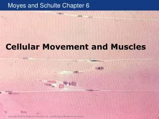
Cellular Movement and Muscles (2). Muscle Cells (Myocytes). Myocytes (muscle cells) Contractile cell unique to animals Contractile elements within myocytes Thick filaments Polymers of myosin ~300 myosin II hexamers Thin filaments Polymers of - actin

E N D
Muscle Cells (Myocytes) • Myocytes (muscle cells) • Contractile cell unique to animals • Contractile elements within myocytes • Thick filaments • Polymers of myosin • ~300 myosin II hexamers • Thin filaments • Polymers of -actin • Ends capped by tropomodulin and CapZ to stabilize • Proteins troponin and tropomyosin on outer surface
Thick and Thin Filaments Figure 5.15
Muscle Cells • Two main types of muscle cells are based on the arrangement of actin and myosin • Striated (striped appearance) • Skeletal and cardiac muscle • Actin and myosin arranged in parallel • Smooth (do not appear striped) • Actin and myosin are not arranged in any particular way
Striated and Smooth Muscle Figure 5.16
Striated Muscle Types Table 5.3
Striated Muscle Cell Structure • Thick and thin filaments arranged into sarcomeres • Repeated in parallel and in series • Side-by-side across myocyte • Causes striated appearance • End-to-end along myocyte
Sarcomeres • Structural features of sarcomeres • Z-disk • Forms border of each sarcomere • Thin filaments are attached to the Z-disk and extend from it towards the middle of the sarcomere • A-band (anisotropic band) • Middle region of sarcomere occupied by thick filaments • I-band (isotropic band) • Located on either side of Z-disk • Occupied by thin filament
Sarcomeres • Thin and thick filaments overlap in two regions of each sarcomere • Each thick filament is surrounded by six thin filaments • Three-dimensional organization of thin and thick filaments is maintained by other proteins • Nebulin • Along length of thin filament • Titin • Keeps thick filament centered in sarcomere • Attaches thick filament to Z-disk
Sarcomeres Figure 5.17
Three-Dimensional Structure of Sarcomere Figure 5.18
Muscle Actinomyosin Activity is Unique • Myosin II cannot drift away from actin • Structure of sarcomere • Duty cycle of myosin II is 0.05 (not 0.5) • Each head is attached for a short time • Does not impede other myosins from pulling the thin filament • Unitary displacement is short • Small amount of filament sliding with each movement of the myosin head
Myofibril • In muscle cells, sarcomeres are arranged into myofibrils • Single, linear continuous stretch of interconnected sarcomeres (i.e., in series) • Extends the length of the muscle cell • Have parallel arrangement in the cell • More myofibrils in parallel can generate more force
Myofibrils in Muscle Cells Figure 5.20
Regulation of Contraction • Excitation-contraction coupling (EC coupling) • Depolarization of the muscle plasma membrane (sarcolemma) • Elevation of intracellular Ca2+ • Contraction • Sliding filaments
Ca2+ Allows Myosin to Bind to Actin • At rest, cytoplasmic [Ca2+] is low • Troponin-tropomyosin cover myosin binding sites on actin • As cytoplasmic [Ca2+] increases • Ca2+ binds to TnC (calcium binding site on troponin) • Troponin-tropomyosin moves, exposing myosin-binding site on actin • Myosin binds to actin and cross-bridge cycle begins • Cycles continue as long as Ca2+ is present • Cell relaxes when the sarcolemma repolarizes and intracellular Ca2+ returns to resting levels
Troponin and Tropomyosin Figure 5.21
Regulation of Contraction by Ca2+ Figure 5.22
Ionic Events in Muscle Contraction Figure 5.23
Troponin–Tropomyosin Isoforms • Properties of isoforms affect contraction • For example, fTnC has a higher affinity for Ca2+ than s/cTnC • Muscle cells with the fTnC isoform respond to smaller increases in cytoplasmic [Ca2+] • Isoforms differ in the affect of temperature and pH
Myosin Isoforms • Properties of isoforms affect contraction • Multiple isoforms of myosin II in muscle • Isoforms can change over time Table 5.4
Excitation of Vertebrate Striated Muscle • Skeletal muscle and cardiac muscle differ in mechanism of excitation and EC coupling • Differences include • Initial cause of depolarization • Time course of the change in membrane potential (action potential) • Propagation of the action potential along the sarcolemma • Cellular origins of Ca2+
Action Potentials • APs along sarcolemma signal contraction • Na+ enters cell when Na+ channels open • Depolarization • Voltage-gated Ca2+ channel open • Increase in cytoplasmic [Ca2+] • Na+ channels close • K+ leave cell when K+ channels open • Repolarization • Reestablishment of ion gradients by Na+/K+ ATPase and Ca2+ ATPase
Time Course of Depolarization Figure 5.24
Initial Cause of Depolarization • Myogenic (“beginning in the muscle”) • Spontaneous • For example, vertebrate heart • Pacemaker cells • Cells that depolarize fastest • Unstable resting membrane potential • Meurogenic (“beginning in the nerve”) • Excited by neurotransmitters from motor nerves • For example, vertebrate skeletal muscle • Can have multiple (tonic) or single (twitch) innervation sites
Neurogenic Muscle Figure 5.25
T-Tubules and Sarcoplasmic Reticulum • Transverse tubules (T-tubules) • Invaginations of sarcolemma • Enhance penetration of action potential into myocyte • More developed in larger, faster twitching muscles • Less developed in cardiac muscle • Sarcoplasmic reticulum (SR) • Stores Ca2+ bound to protein sequestrin • Terminal cisternae increase storage • T-tubules and terminal cisternae are adjacent to one another
T-Tubules and SR Figure 5.28
Ca2+ Channels and Transporters • Channels allow Ca2+ to enter cytoplasm • Ca2+ channels in cell membrane • Dihydropyridine receptor (DHPR) • Ca2+ channels in the SR membrane • Ryanodine receptor (RyR) • Transporters remove Ca2+ from cytoplasm • Ca2+ transporters in cell membrane • Ca2+ ATPase • Na+/Ca2+ exchanger (NaCaX) • Ca2+ transporters in SR membrane • Ca2+ ATPase (SERCA)
Ca2+ Channels and Transporters Figure 5.27
Induction of Ca2+ Release From SR • AP along sarcolemma conducted down T-tubules • Depolarization opens DHPR • Ca2+ enters cell from extracellular fluid • In heart, [Ca2+] causes RyR to open, allowing release of Ca2+ from SR • “Ca2+ induced Ca2+ release” • In skeletal muscle, change in DHPR shape causes RyR to open, allowing release of Ca2+ from SR • “Depolarization induced Ca2+ release”
Ca2+ Induced Ca2+ Release Figure 5.29
Depolarization Induced Ca2+ Release Figure 5.30
Relaxation • Repolarization of sarcolemma • Remove Ca2+ from cytoplasm • Ca2+ ATPase in sarcolemma and SR • Na+/Ca2+ exchanger (NaCaX) in sarcolemma • Parvalbumin • Cytosolic Ca2+ binding protein buffers Ca2+ • Ca2+ dissociates from troponin • Tropomyosin blocks myosin binding sites • Myosin can no longer bind to actin
Relaxation Figure 5.27
Summary of Striated Muscles Table 5.5