Download
1 / 17
170 likes | 382 Views
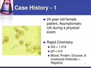
Case History. 67 yo F Progressive visual loss in the SO associated with corneal degeneration and a limbal tumor Gross description Opaque white tissue measures 13x12mm Extends from the limbus to the edge of cornea. Diagnosis Dysplasia/CIN, limbal conjunctiva.

E N D
Case History • 67 yo F Progressive visual loss in the SO associated with corneal degeneration and a limbal tumor • Gross description • Opaque white tissue measures 13x12mm • Extends from the limbus to the edge of cornea
Diagnosis • Dysplasia/CIN, limbal conjunctiva
Conjunctival intraepithelial neoplasm (CIN) • Definition: CIN includes a wide range of neoplastic intraepithelial changes ranging from dysplasia to full-thickness epithelial neoplasia or carcinoma in situ • Synonyms include mild, moderate, and severe dysplasia, carcinoma in situ, ocular surface squamous neoplasia, intraepithelioma and bowenoid dyskeratosis
CIN • Clinically, lesions are sharply demarcated and arise at the limbus, where corneal stem cells are located, with either or both conjunctival and corneal involvement. • Most lesions are pink, nonkeratinized, well- vascularized and have a raspberry-like configuration. • Rarely, these changes spontaneously regress.
CIN • Microscopic findings • Keratinization and dyskeratosis are not a common feature of CIN. • Atypical mitosis are frequent and may be located at all levels of the epithelium. • The intraepithelium dysplastic changes are graded as mild, moderate, or severe based on the thickness of intraepithelial involvement.
CIN • Differential diagnosis. • UV-related epithelial hyperplasia • Intraepithelial sebaceous gland carcinoma • Inraepithelial invasion by adenocarcinoma originating from aprocrine glands of moll • Primary acquired melanosis (HMB45, S-100, Melan A)
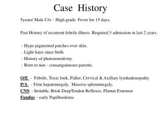
Case History • 50 yo F • 2Y/H progressively enlarging pigmented area involving the conjunctiva and cornea of the left eye. • 3X3.5 mm. pigmented limbal nodule appeared in this area of heavily pigmented conjunctiva, which had been previously biopsied and diagnosed as “primary acquired melanosis”
Case History (Cont.) • The left eye and a broad zone of bulbar conjunctiva were excised because of the clinical diagnosis of malignant melanoma of the conjunctiva.
Diagnosis • Malignant melanoma of limbus arising from primary acquired melanosis.
Melanocytic tumors of the conjunctiva • Ephelis(Freckle) • Lentigo • Nevus • Primary Acquired Melanosis(PAM) • Malignant Melanoma
PAM • Clinical Characteristics • The melanosis of unilateral, diffuse, brown pigmentation that moves with the conjunctiva over the sclera (analogous to lentigo maligna of the skin). • Age of onset is 40 to 50 years of age. • No clinical differentiation in PAM with or without atypia
Case History • 60 yo M • H/O Pseudophakic bullous keratopathy • Waxing and waning corneal edema • Decreased vision in his left eye • Penetrating keratoplasty