Download
1 / 43
430 likes | 505 Views
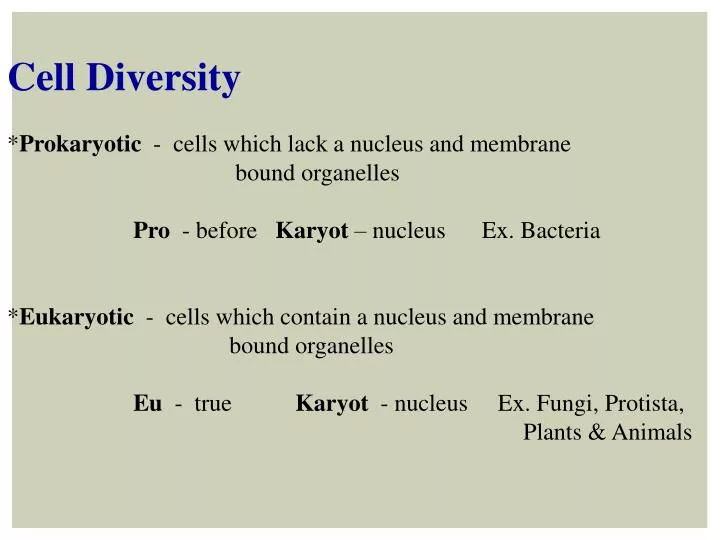
Cell Diversity * Prokaryotic - cells which lack a nucleus and membrane bound organelles Pro - before Karyot – nucleus Ex. Bacteria * Eukaryotic - cells which contain a nucleus and membrane

E N D
Cell Diversity *Prokaryotic - cells which lack a nucleus and membrane bound organelles Pro - before Karyot– nucleus Ex. Bacteria *Eukaryotic - cells which contain a nucleus and membrane bound organelles Eu - true Karyot - nucleus Ex. Fungi, Protista, Plants & Animals
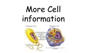
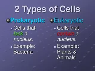
Prokaryotic vs. Eukaryotic • Prokaryotic cells….. • do not enclose their genetic material within a • nucleus • are generally smaller and simpler then eukaryotic cells - always unicellular • do not have membrane-bound organelles • DNA is circular • Ribosomes are small • Moves by rotating flagellum • Cell division by binary fission
Escherichiacoli Staphylococcusepidermidis
Prokaryotic vs. Eukaryotic • Eukaryoticcells….. • use their nucleus to enclose the genetic material from the rest of the cell • are generally larger and more complex then prokaryotic cells –multicellular • have membrane-bound organelles • DNA is linear • Ribosomes are large • Moves by waving cilia or flagellae • Cell division by mitosis or meiosis
Prokaryotic & Eukaryotic Similarities: • Enclosed by plasma membranes • Contain DNA as their genetic material • Have Ribosomes • Filled with cytoplasm
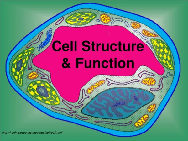
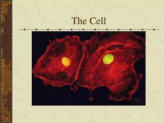
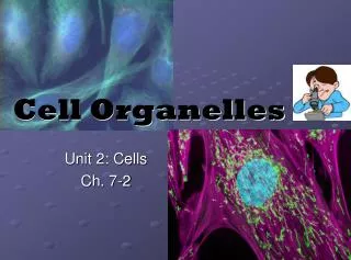
Eukaryotic Organelles “ little organs” We will spend the next section discussing the different organelles found in Eukaryotic Cells Organelles found in a cell are dependent on the cell’s particular function in the organism. All cells have organelles which are necessary for the cells own survival needs like: *Food intake *Conversion of food to Cellular Energy *Waste Removal *Reproduction
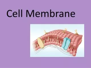
*No matter how cells differ from one another the majority of Eukaryotic cells have these Three Main Components: 1. Cell membrane- outer boundary of the cell 2. Cytoplasm - liquid portion inside the cell membrane 3. Nucleus - contains the cells DNA 1. Cell Membrane *The cell membrane is also known as the Plasma Membrane. A. Functions: 1. Separates the cell from its external surroundings or environment 2. Gives the cell its shape and flexibility
3. Regulates what enters and exits the cell. • Selectively permeable/ Semi-permeable • *to permeate - to pass through
2. Cytoplasm *Also called Cytosol - a gelatin-like aqueous fluid which bathes the organelles which is found between the cell membrane & the nucleus *Aqueous fluid which contains: Salt(0.85%) Organic molecules (? Proteins, Carbohydrates + Lipids) Minerals (K Mg Na Fe ) *The cytoplasm is in constant motion as particles and organelles move around inside the cell called Cytoplasmic Streaming
3. Nucleus A. Function: * directs cellular activity * contains most of the cells DNA * site of nucleic acid synthesis ( RNA & DNA) B. Structure: * spherical organelle with thousands of nuclear pores * surrounded by a double membrane =Nuclear Envelope *contains a spherical body called the Nucleolus, where ribosomes are produced * it is porous, so small substances can pass in and out *filled w/ a dense liquid =Nucleoplasm
4. Ribosomes A. Function: * site of Protein Synthesis *most abundant organelle in the cell B. Structure: *consists of 2 spheres (upside down snowman) * smallest organelle (15 - 20 nm) *not membrane bound *made in the nucleus by the nucleolus
C. Location: * 2 locations of Ribosomes: 1. Free Floating in the Cytoplasm - makes proteins for the Cell’s Own Use, to be used inside the cell Ex. Cellular Enzymes 2. Attached to the Endoplasmic Reticulum - makes proteins to be exported from the cell to be used in other areas of the organism Ex. Insulin Gastric Enzymes
5. Endoplasmic Reticulum (ER) A. Structure: *a system of highly folded membranes forming pouches and tunnels *lipid/proteins components are assembled B. Functions: *the function of the endoplasmic reticulum depends on whether it has ribosomes attached or not. 2 Types of ER: 1. Rough ER - covered with ribosomes resulting in a bumpy or rough appearance a. site of synthesis of exported proteins b. intracellular pathway
2. Smooth ER - has no ribosomes on its surface so it appears smooth a. intracellular pathway b. storage area for proteins waiting to be exported c. synthesis of steroids in Gland Cells d. Regulates Ca levels in Muscle Cells e. Breakdown of toxic substances in Liver Cells 6. Golgi Apparatus *also called Golgi Bodies A. Structure: *a stack of membranes and fluid filled sacs *fluid contains dissolved substances to be added to proteins before they are exported
B. Function: * processing packaging and secreting of proteins * like an assembly line in a manufacturing plant: 1. Proteins are made in Rough ER *Car gets body, wheels, frame etc. 2. Travel through the Rough ER and /or Smooth ER *Assembly line 3. Placed in a membrane vesicle *Car carrier takes the car to the next station 4. Travels to the Golgi to get the extras *Options are added: CD, AC, Navigation etc 5. Placed into another vesicle *Another car carrier 6. Travels to the Cell Membrane to be exported *Out the factory doors
7. Vesicles A. Function: *Stores or transports substances within a cell *most have specialized functions depending on what material they contain B. Structure: *Small, intracellular membrane-enclosed sac
8. Mitochondria A. Function: *the power house of the cell *where compounds are broken down through the process of Cellular respiration B. Structure: *a large organelle composed of 2 membranes: 1. Outer Membrane - separates the mitochondria from its surroundings 2. Inner membrane - highly folded membrane across which respiration takes place called the Cristae
What is the advantage of having the Cristae highly folded? > Surface Area, > amount of energy produced What type of cells have high numbers of Mitochondria? Muscle Cells 2500/cell *Theory of Endosymbiosis: * Notice the shape of the Mitochondria. Does it remind you of any cells we have discussed before? 1. Scientists believe that mitochondria were once rod shaped Prokaryotic Bacteria that were engulfed by other bacteria. 2. The inner bacteria provided energy and the outer cell provided protection. 3. This was a Symbiotic Relationship in which both bacteria benefited . 4. Over time they became so dependent on each other that they could not live apart and developed into the first Eukaryotic Cells.
9. Lysosomes A. Structure: * small spherical organelles which contain over 40 digestive enzymes B. Function: * digest food particles like: carbohydrates, proteins, lipids, DNA & RNA into their monomers so cell can use them for energy or reuse them to make molecules needed * also digests foreign invaders like Viruses and Bacteria to protect the cell
10. Cytoskeleton, Microtubules & Microfilaments A. Function: *Cells need a support system = called a Cytoskeleton *The cytoskeleton helps the cell maintain its shape and is involved in moving “spider web-like” B. Structure: *There are 2 types of proteins involved: 1. Microtubules - long hollow tubes made of tubulin (protein) used to make Spindle fibers which pull apart chromosomes during cell division
2. Microfilaments - threadlike structures made of actin (protein) their assembly and disassembly are responsible for cytoplasmic streaming
11. Centrioles A. Function: *help to organize cell division B. Structure:*located near nucleus, made from the protein tubulin * not in plants
12. Cilia & Flagella A. Function: *responsible for cellular movement 1. Cilia - short hair-like structures which extend from the cell *usually in high numbers *surround the cell *beat in synchronized strokes
Functions of Cilia: Unicellular organisms a. locomotion Ex. paramecium Multicellular organisms a. move substances along the digestive tract b. filter out dirt and debris in the respiratory tract Ex. Nose 2. Flagella - long whip-like structures *usually found in singles or pairs Functions of Flagella: Unicellular organisms a. locomotion Ex. Giardialamblia Multicellular organisms a. locomotion Ex. sperm
The Next 3 Organelles are Found Primarily in Plants 13. Cell Wall A. Function: * a rigid covering that surrounds the cell membrane which is inflexible B. Structure: * made primarily of a polysaccharide =Cellulose * contains pores to allow substances to enter which are then regulated by the semi-permeable cell membrane
14. Central Vacuole A. Structure: *a fluid filled space located in the center of a plant cell *can take up as much as 90% of the cells volume. B. Function *stores enzymes and metabolic wastes (The plant needs to eliminate its waste but the cell wall limits this. By placing the metabolic waste in the vacuole with enzymes, the waste products can be broken down into usable substances) *location of plant poisons ****There are vacuoles in animal cells too!! -Smaller, sac-like membrane enclosed structures -Store materials like water, salts, proteins and carbs -Play a role in the release of cellular waste
15. Chloroplast A. Structure: * green organelle which contains the pigment Chlorophyll * contains flattened sacs called thylakoids B. Function: * where photosynthesis takes place
Escherichiacoli Staphylococcusepidermidis
Onion Elodea Human Cheek
Gram staining is a microbiological procedure that categorizes bacteria based on the physical and chemical structure of their outer surface. Gram-positive bacteriahave a thick layer made up of polymers of protein-sugar molecules called peptidoglycan. Gram staining of the peptidoglycan layer (a.k.a. the cell wall) with a chemical called crystal violet results in purple coloration of the gram-positive bacteria. Addition of acetone or alcohol dehydrates the bacteria, causing it to retain the purple color. Gram-negative bacteriahave an additional outer membrane whose properties resist acetone/alcohol-dehydration and result in loss of the crystal violet stain. A counterstain, made up of fuchsin, stains these bacteria red or pink.