Download
1 / 12
120 likes | 340 Views
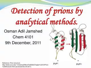
Detection of prions by analytical methods. Osman Adil Jamshed Chem 4101 9th December, 2011. Prions. Prions are misfolded form of proteins classified as infectious pathogens.

E N D
Detection of prions by analytical methods. Osman Adil Jamshed Chem 4101 9th December, 2011
Prions • Prions are misfolded form of proteins classified as infectious pathogens. • Responsible for fatal neurodegenerative diseases in mammals by modifying cellular prion protein (PrPc) to infected scarpieprion protein (PrPSc). • Transmissible spongiform encephalopathies – the transmissible form of prion diseases. • Creutzfeldt – Jakob disease (CJD) in humans, Bovine spongiform encephalopathy (BSE) in cattle, Scarpie of sheep.
Mechanism of Prion conversion. Structure 'a' is normal PrP with only alpha helix and structure 'b' is prion with beta sheets Possible mechanism for the conversion.
Problem • Extremely stable and resistant to chemical and physical agents like heat, acids, alkalis, detergents and enzymatic proteolysis. • A “slow virus” as it kills human over a period of several years. • The disease fatal and virtually undetectable until its too late. • Simple cooking of foodborne variant Creutzfeldt-Jakob disease infected meat products especially brain, does not stop the transmission. • CJD is a variant of BSE. Animals products used in making feed in some countries like UK. • The protein isoforms are quite similar to each other makes detection hard.
Hypothesis and Studies • Preventing the spread the prions in animal feed product would prevent a future BSE epidemic and prevent infecting humans with vCJD – a variant of BSE. • The studies would include analyzing animal by-productsmade from them, in regions (factories nearby) with reported prion outbreaks and places with no prion out break. The data will be compared to the safety precautions taken by factories in the region. • Analyte = PrPSc; Matrices = biological, brain and CNS tissue.
Q-TOF mass spectrometer operating in MS (upper) and MS/MS mode (lower) modes.
Sample preparation • Brain or nervous tissue will be homogenized (HT1000 Potter homogenizer) in ice-cold PBS containing 0.5% Nonidet p-40 and 0.5% deoxycholate to give a 10% (w/v) final suspension. • Homogenates will be centrifuged for 10 min at 5000 xg at 4 oC. • Supernatants of brain or nervous tissue homogenates were aliquoted and stored at -80 oC. • Before analysis, bovine trysin (3.3μg in 33 μL water) digestion will be carried out at 37 oC overnight. • The digestion will be stopped by adding formic acid (2μL) to give a pH of 2.3.
Method specifications • Nanospray LC/ ESI/ Q-TOF • Applied Biosystems MDS SCIEX QStar Pulsar equipped with ProxeonBiosystemsnanoelectrospray source schematics (left diagram) and ESI with MS (right one) shown below. MS/MS real life picture ESI process Q-TOF MS/MS
Conclusion • The nanoLC/MS/MS was considered to be the best technique due to high selectivity and it provides high sensitivity for qualitative measurements. • MS/MS can detect the prions in biological matrices in sub-femtomole amounts. • The sensitivity can be increased by the use of phenylisothiocyanate (PITC) derivatives.
References • http://www.fda.gov/Food/FoodSafety/FoodborneIllness/FoodborneIllnessFoodbornePathogensNaturalToxins/BadBugBook/ucm071397.htm • Prusiner, S.; Proc. Natl. Acad. Sci.,1998, 95, pp. 13363–13383 • Shkundina, I. S.; Ter-Avanesyan, M. D.; Biochemistry (Moscow), 2007, 72, 13, pp. 1519-1536 • Onisko, B.; Dynin, I.; Requena, J.; Silva, C.; Erickson, M.; Carter, J.; J. Am. Soc. Mass Spectrom., 2007, 18, 1070-1079. • Ramsay, L.; Dickerson, J.; Dovichi, N.; Electrophoresis 2009, 30, 297–302 • Serbec, V.; Bresjanac, M.; Popovic, M.; et al, J. Bio. Chem. 2004, 279, 5, 3694-3698. • http://www.spectroscopynow.com/coi/cda/detail.cda?id=11316&type=EducationFeature&chId=10&page=1 • http://www.unige.ch/sciences/sms/2.html • http://www.tau.ac.il/lifesci/units/proteomics/qstar.html • http://www.colorado.edu/chemistry/chem5181/QSTAR%20Pulsar.pdf • Wilham, J.; Orru, C.; Bessen, R.; Atarashi, R.; Sano, K.; Race, B.; Meade-White, K.; Taubner, L.; Timmes, A.; Caughey, B.; PLoS Pathogens, 2010, 6, 12, pg 1-15