Download
1 / 46
470 likes | 626 Views
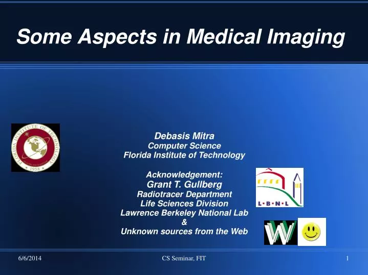
Some Aspects in Medical Imaging. Debasis Mitra Computer Science Florida Institute of Technology Acknowledgement: Grant T. Gullberg Radiotracer Department Life Sciences Division Lawrence Berkeley National Lab & Unknown sources from the Web. Co-ordinates. Why this talk? Where am I now?

E N D
CS Seminar, FIT Some Aspects in Medical Imaging Debasis Mitra Computer Science Florida Institute of Technology Acknowledgement: Grant T. Gullberg Radiotracer Department Life Sciences Division Lawrence Berkeley National Lab & Unknown sources from the Web
CS Seminar, FIT Co-ordinates • Why this talk? • Where am I now? • What does this lab do?
CS Seminar, FIT Lawrence Berkeley National Lab
CS Seminar, FIT Center for Functional Imaging
CS Seminar, FIT Biomedical Imaging is the Engineering behind Radiology
CS Seminar, FIT Types of Imaging Instruments • Computer Tomography (X-ray) • Magnetic Resonance Imaging (MRI) • Single Photon Emission Computed Tomography (SPECT): gamma ray of 100-few hundred kev • Positron Emission Tomography (PET): gamma ray from in situ positron annihilation, 500 kev • Ultra Sound • Optical or Laser Tomography (Infrared) • Fluoroscopy, Opto-acoustic, Electron, Atomic-force, Radio-frequency,…
CS Seminar, FIT Resolution Sensitivity GE VG3 Millennium Hawkeye SPECT/CT collimators -ray detectors x,y Acquisition system
CS Seminar, FIT Scintillation Camera and Collimator Patient Collimator localizes events in object and determines sensitivity and spatial resolution of the camera
CS Seminar, FIT Collimator converging parallel hole pinhole
CS Seminar, FIT Positron Emission Tomography Does Not Need a Collimator • Positron annihilates with electron two gamma photons each at 511 keV leave under 180 • Coincidence detection (“electronic collimation”)
CS Seminar, FIT PET
CS Seminar, FIT MRI Epilepsy: MRI, PET-time 1, 2 Brain tumor
CS Seminar, FIT A B C D E F Fiber Tracking of DTMRI Data Rohmer D, Sitek A, Gullberg GT: Reconstruction and visualization of fiber and laminar structure in the normal human heart from ex vivo DTMRI data. Investigative Radiology, 42:777-789, 2007.
CS Seminar, FIT Ultrasound
CS Seminar, FIT CardiARC
CS Seminar, FIT Clinical FeasibilityResults Spectrum Dynamics Conventional 1.45 Mcounts total (heart 10%, backgnd 90%) Pixel size 6.91 mm × 6.91 mm Iterative reconstruction Total acquisition time: 17.5 min 0.8 Mcounts total (heart 60%, backgnd 40%) Pixel size 2.46 mm × 6.91 mm × 6.91 mm Iterative reconstruction Total acquisition time: 2.2 min
CS Seminar, FIT Radiopharmaceuticals forCardiac Imaging • 201Tl • 99mTc-sestamibi (2-Meth0xy-2-methylpropyl isonitrile) • 99mTc-tetrafosmin • 99mTc-teboroxime • 123I-iodorotenone • 123I-BMIPP (fatty acid) • 123I-IPPA (fatty acid)
CS Seminar, FIT Targets of Study • Heart, • Lungs, liver, other organs in torso • Brain: Alzheimer’s Disease Neuroimaging Initiative (ADNI) • Breast • Tumor Breast Cancer
CS Seminar, FIT Physics behind Models • Emission tomography: SPECT, PET, MRI • Transmission tomography: X-ray, Optical • Reflection: Ultra Sound, Total Internal • Reflection Fluoroscopy (TIRF for single cell • visualization) • Scattering: Muontomography?
CS Seminar, FIT Mathematical Problem Formulation • Forward Problem (modeling): How the data would look like • given probe and the model • D = F(M): Forward project • An implementation is a Simulation software • Inverse Problem (tomography): What the model would be • given the probe and data • M = F ~ (D): back-project • An implementation is a Reconstruction software • Noise in data makes it a hard statistical problem • Data volumemay be additional computational • challenge • http://en.wikipedia.org/wiki/Inverse_problem
CS Seminar, FIT Reconstruction Algorithms • Analytic-inverse: E.g., Radon transformation for • emission/absorption (mostly useless except for theoretical • purpose) • Algebraic Reconstruction: voxel by voxel reconstruct the model • Iterative Reconstruction using Expectation Maximization • Ordered Set – EM • Maximum A Posteriori (MAP-EM) • Penalized Least Square (PLS): 1.5 iteration!
CS Seminar, FIT Dynamic Imaging • Problem: Objects move during data gathering • Question: How to reconstruct (1) Object, (2) Motion • A successful approach: Level Set • For blood concentration change in tissues: • Temporal B-spline • Tensor imaging with MRI
CS Seminar, FIT k32 k21 C2(t) C3(t) blood k23 k12 Fit the 123I-BMIPP Data to a Compartment Model • Need to estimate an input function. • Time activity curves have to be estimated directly from the projections. • A methyl group on the position of the carbon chain limits the oxidation of 123I-BMIPP. • Differs from 123IPPA which is completely metabolized to benzoic acid. TG IPPA Model of IPPA Metabolism Benzoic acid
CS Seminar, FIT Spatiotemporal Modeling Using A Small Number of Splines to Represent Realistic Physiological Curves • Quadratic B-Spline Temporal Basis Functions • Zero Order (voxels) B-Spline Spatial Bases
CS Seminar, FIT 0o 8o 16o 24o 32o 40o 48o 56o 64o 80o 72o 88o 96o 104o 112o 120o 128o 136o 352o Slow-Rotation Dynamic Pinhole SPECT Blood Time Activity Curve Estimated from Projections Using Factor Analysis 1 sec frames, 180˚ rotation of one head Recirculation time is 6-8 seconds
CS Seminar, FIT Results — Dynamic Early Data
Pixels / voxels Blobs Linear B-splines Cubic B-splines “Custom-made” shapes Irregular meshes . . . . . . . . . . . . . . Image Spatial Representations regular sparse
CS Seminar, FIT Metabolic Rate of BMIPP Normal Ki=0.40 min-1 SHR Ki=0.15 min-1
CS Seminar, FIT Metabolic Rate: FDG vs BMIPP BMIPP FDG Ki=0.40 min-1 Ki=0.15 min-1
CS Seminar, FIT WKY: normal rat
Flow rate changesSHR: Hypertensive, WKY: normal CS Seminar, FIT
CS Seminar, FIT SHR red A C A B 0.90 0.60 0.00 0.60 B Normal 6/18/2003 8/06/2003 10/01/2003 12/2/2003 7/14/2004 9/21/2004 0.60 0.30 SHR red 0.00 C 0.00 6/18/2003 8/06/2003 10/01/2003 4/27/2004 1.25 Forward Warping 0.85 D Normal 6/18/2003 8/06/2003 10/01/2003 12/2/2003 7/14/2004 9/21/2004 1.25 0.85 6/18/2003 8/06/2003 10/01/2003 4/27/2004 Temporal Comparison of 1st Principal Strain for SHR and WKY anterior wall septum SHR 1st PS WKY FS Veress A et al.: Regional changes in the diastolic deformation of the left ventricle for SHR and WKY rats using 18FDG based microPET technology and hyperelastic warping. Annals of Biomedical Engineering 36:1104–1117, 2008.
CS Seminar, FIT Parametric Imaging Summed Images (between 2 and 12 min) Parametric Images of k21 Sitek A, Di Bella EVR, Gullberg GT, Huesman RH: Removal of liver activity contamination in teboroxime dynamic cardiac SPECT imaging using factor analysis. J Nucl Cardiology 9:197-205, 2002.
CS Seminar, FIT SUMMARY • The SHR shows increased glucose metabolism and reduced fatty acid metabolism. • The reverse is true for the nomotensive WKY rat. • The SHR model is used to develop techniques for analysis of imaging data of heart failure related to metabolism. • Molecular Insight Pharmaceuticals is now evaluating 123I-BMIPP in clinical trials. • These results of fatty acid metabolism correlate with those in humans with hypertensive left ventricular hypertrophy.(de las Fuentes et al. J NuclCardiol 13:369, 2006)
CS Seminar, FIT COMMENTS • The SHR has a defective gene (CD36) on chromosome 4. • The defect is associated with compromised long-chain FA transport across the cell membrane. • The defect causes insulin resistance, alteration in basal glucose metabolism. • Short-chain FA diet decreases glucose uptake, alleviates hypertrophy, but hypertension is not improved. • Proposed research will compare 123I-BMIPP with 18FTHA. Hajri T et al.: Defective fatty acid uptake in the spontaneously hypertensive rat is a primary determinant of altered glucose metabolism, hyperinsulinemia, and myocardial hypertrophy. J Biological Chem 276:23661-23666, 2001.
MRI is way advanced in Dynamic Imaging Diffusion Tensor Imaging A high-resolution diffusion tensor imaging scan reveals differences between healthy tracts of axons, at left and in the lower enlargement, and tracts of injured axons, at right and in the top enlargement, in a person who sustained a moderate to severe traumatic brain injury. Such damage has been shown to correlate with cognitive impairment. (Image courtesy of Dr. Deborah Little) CS Seminar, FIT
CS Seminar, FIT Diffusion Tensor Diffusion within a single voxel. (a) Diagram shows the 3D diffusion probability density function in a voxel that contains spherical cells (top left) or randomly oriented tubular structures that intersect, such as axons (bottom left). This 3D displacement distribution, which is roughly bell shaped, results in a symmetric image (center), as there is no preferential direction of diffusion. The distribution is similar to that in unrestricted diffusion but narrower because there are barriers that hinder molecular displacement. The center of the image (origin of the r vector) codes for the proportion of molecules that were not displaced during the diffusion time interval.
CS Seminar, FIT A B C D F E Sheet Tracking of DTMRI Data
CS Seminar, FIT B A Fiber Tracking of Right and Left Ventricle Cardiac Band Hypothesis: The four chamber heart is built from a single continuous band of muscle. Torrent-Guasp F, Kocicab MJ, Cornoc AF, et al. Towards new understanding of the heart structure and function. Eur J Cardiothorac Surg. 2005;27:191-201.
CS Seminar, FIT Advancement of Data Acquisition Technology • List mode: acquire data for recording time for each • track and reconstruct with it: a computational challenge • Time-of-flight: Acquire event versus data collecting • time: new type of detectors needed • Compton gamma camera: provides some measure of • angle of a track • Newer Technology: Opto-acoustic, Fluorescence, … • Target-specific detectors: e.g., Cardiac-Spect, faster • and cleaner data with higher resolution
CS Seminar, FIT Molecular Imaging • Medical imaging is primarily at organ-level • With more genetic information available today it is usual to think in terms of metabolism behind images, and target cellular-level processes • Current focus is to develop ligands that are • tagged with imaging agents, (2) binds to some protein or metabolite that we want to visualize with imaging • Understanding dynamic organ-level images from metabolic point of view is another new area
CS Seminar, FIT Total Internal Reflection Imaging TIRF imaging of actin networks and their reorganization in the cortex of Dictyostelium cells.
CS Seminar, FIT Auto-diagnosis/prognosis:Machine learning • Images are still used by radiologists for • diagnosis/prognosis, or by biologist for doing science: • technology targets exclusively to improve • image quality, and nothing more • It is quite possible to use machine learning algorithms • to help the process: • image is input, zones of interest with annotations are output
CS Seminar, FIT Thanks! Debasis Mitra dmitra@cs.fit.edu