Download
1 / 11
140 likes | 322 Views
Special Circulations. Arterial supply of the brain and the circle of Willis. The brain is supplied by two pairs of arteries, the internal carotid arteries and the vertebral arteries

E N D
Arterial supply of the brain and the circle of Willis • The brain is supplied by two pairs of arteries, the internal carotid arteries and the vertebral arteries • Internal carotid arteries: branches of the common carotid arteries, run through the neck and enter the skull through the temporal bone. • Once inside the cranium, each divides into the anterior and middle cerebral arteries, which supply most of the cerebrum
Arterial supply of the brain and the circle of Willis • The paired vertebral arteries: It pass upward from the subclavian arteries at the base of the neck. • Within the skull, the vertebral arteries join to form the single basilar artery, which serves the brain stem and cerebellum as it travels upward • At the base of the cerebrum, the basilar artery divides to form the posterior cerebral arteries, which supply the posterior part of the cerebrum
Arterial supply of the brain and the circle of Willis • The anterior and posterior blood supplies of the brain are united by small communication arterial branches • The result is a complete circle of connecting blood vessels called the circle of Willis, which surround the base of the brain, because it provides more than one route for blood to reach brain tissue
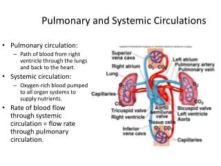
Hepatic Portal Circulation • The veins of the hepatic portal circulation drain the digestive organs, spleen and pancreas, and deliver this blood to the liver through the hepatic portal vein • As blood flows through the liver, some of the nutrients are removed to be stored or processed in various ways for later release to the blood
Hepatic Portal Circulation • The inferior mesenteric vein, draining the terminal part of the large intestine, drains into the splenic vein, which itself drains the spleen, pancreas, and the left side of the stomach • The splenic vein and superior mesenteric vein join to form the hepatic portal vein • The gastric vein, which drains the right side of the stomach, drains directly into the hepatic portal vein
Fetal Circulation • Since the lungs and digestive system are not yet functioning in a fetus, all nutrient, excretory and gas exchange occur thorough the placenta • The umbilical cord contains 3 blood vessels: • Large umbilical vein • 2 smaller umbilical arteries
Fetal Circulation • The umbilical vein carries blood rich in nutrients and oxygen to the fetus • The umbilical arteries carry carbon dioxide from the fetus to the placenta
Fetal Circulation • As blood flows superiorly toward the heart of the fetus, most of it by passes the immature liver through the ductus venous and enters the inferior vena cava • Since fetal lungs are nonfunctional and collapsed, they are almost entirely bypassed
Fetal Circulation • Some blood entering the right atrium is directed into the left atrium through a flap like opening in the interatrial septum, the foramen ovale • Blood that does manage to enter the right ventricle is pumped out the pulmonary trunk, where it meets the ductus arteriosus, a short vessel that connects the aorta and the pulmonary trunk Foramen ovale Ductus arteriosus
Fetal Circulation • At birth, the foramen ovale closed and the ductus arteriosus collapses and is converted to fibrous ligamentum arteriosum • As blood stops flowing through the umbilical vessels, they obliterated, and the circulator pattern becomes that of an adult